Abstract
Purpose
To evaluate the clinical outcomes of implantable collamer lens (ICL) implantation with simultaneous full thickness as-tigmatic keratotomy (FTAK) for the correction of moderate to high myopic astigmatism.
Methods
Thirty-two eyes of 16 patients who had an ICL implantation with simultaneous FTAK were studied. Follow-up visits were at 1 week, 1 month, and 3 months. The outcome measures included the uncorrected distance visual acuity (UDVA), re-fractive error, efficacy, safety, and predictability.
Results
After the surgery, astigmatism was reduced by 74.83 ± 13.8%. The proportion of eyes with a spherical equivalent of 0.5 D or less was 87.5%, and all eyes had a spherical equivalent of 1.0 D or less at 3 months after the surgery. The proportion of eyes with a UDVA of 20/25 or better was 100%, and 20/20 or better was 81.25%. Reoperation was needed in one case (3.1%) because of undercorrection of the astigmatism, and no complications were observed.
Go to : 
References
1. Kamiya K, Shimizu K, Igarashi A. . Four-year follow-up of posterior chamber phakic intraocular lens implantation for moder-ate to high myopia. Arch Ophthalmol. 2009; 127:845–50.

2. Alfonso JF, Baamonde B, Fernández-Vega L. . Posterior cham-ber collagen copolymer phakic intraocular lenses to correct my-opia: five-year follow-up. J Cataract Refract Surg. 2011; 37:873–80.

3. Igarashi A, Shimizu K, Kamiya K. Eight-year follow-up of posteri-or chamber phakic intraocular lens implantation for moderate to high myopia. Am J Ophthalmol. 2014; 157:532–9.e1.

4. Zaldivar R, Davidorf JM, Oscherow S. Posterior chamber phakic intraocular lens for myopia of-8 to- 19 diopters. J Refract Surg. 1998; 14:294–305.
5. Alfonso JF, Palacios A, Montés-Micó R. Myopic phakic STAAR collamer posterior chamber intraocular lenses for keratoconus. J Refract Surg. 2008; 24:867–74.

6. Kamiya K, Shimizu K, Aizawa D. . One-year follow-up of pos-terior chamber toric phakic intraocular lens implantation for mod-erate to high myopic astigmatism. Ophthalmology. 2010; 117:2287–94.

7. Kamiya K, Shimizu K, Kobashi H. . Three-year follow-up of posterior chamber toric phakic intraocular lens implantation for moderate to high myopic astigmatism. PLoS One. 2013; 8:e56453.

8. Yoon JM, Moon SJ, Lee KH. Clinical outcomes of toric implant-able collamer lens implantation. J Korean Ophthalmol Soc. 2009; 50:839–51.

9. Kim BK, Mun SJ, Lee DG, Chung YT. Clinical outcomes of com-bined procedure of astigmatic keratotomy and laser in situ keratomileusis. J Korean Ophthalmol Soc. 2016; 57:353–60.
10. Kim BK, Mun SJ, Lee DG. . Full-thickness astigmatic keratot-omy combined with small-incision lenticule extraction to treat high-level and mixed astigmatism. Cornea. 2015; 34:1582–7.

11. Kim BK, Mun SJ, Lee DG, Chung YT. Clinical outcomes of bev-eled, full thickness astigmatic keratotomy. J Korean Ophthalmol Soc. 2015; 56:1160–9.

12. Tatar MG, Aylin Kantarci F, Yildirim A. . Risk factors in post-LASIK corneal ectasia. J Ophthalmol. 2014; 2014:204191.

13. Ivarsen A, Næser K, Hjortdal J. Laser in situ keratomileusis for high astigmatism in myopic and hyperopic eyes. J Cataract Refract Surg. 2013; 39:74–80.

14. Bhandari V, Karandikar S, Reddy JK, Relekar K. Implantable col-lamer lens V4b and V4c for correction of high myopia. J Curr Ophthalmol. 2016; 27:76–81.

15. Park SC, Kwun YK, Chung ES. . Postoperative astigmatism and axis stability after implantation of the STAAR toric implant-able collamer lens. J Refract Surg. 2009; 25:403–9.

16. Mori T, Yokoyama S, Kojima T. . Factors affecting rotation of a posterior chamber collagen copolymer toric phakic intraocular lens. J Cataract Refract Surg. 2012; 38:568–73.

17. Gomez-Bastar A, Jaimes M, Graue-Hernández EO. . Long- term refractive outcomes of posterior chamber phakic (spheric and toric implantable collamer lens) intraocular lens implantation. Int Ophthalmol. 2014; 34:583–90.
18. Kamiya K, Shimizu K, Aizawa D. . Surgically induced astig-matism after posterior chamber phakic intraocular lens implantation. Br J Ophthalmol. 2009; 93:1648–51.

19. Pineda R, Jain V. Arcuate keratotomy: an option for astigmatism correction after laser in situ keratomileusis. Cornea. 2009; 28:1178–80.

20. Song HB, Choi HJ, Kim MK, Wee WR. The short-term effect of limbal relaxing incision and compression suture on post-penetrating keratoplasty astigmatism. J Korean Ophthalmol Soc. 2011; 52:1142–9.

21. Nubile M, Carpineto P, Lanzini M. . Femtosecond laser arcuate keratotomy for the correction of high astigmatism after keratoplasty. Ophthalmology. 2009; 116:1083–92.

22. Hoffart L, Proust H, Matonti F. . Correction of postkeratoplasty astigmatism by femtosecond laser compared with mechanized as-tigmatic keratotomy. Am J Ophthalmol. 2009; 147:779–87.e1.

23. Buzzonetti L, Petrocelli G, Laborante A. . Arcuate keratotomy for high postoperative keratoplasty astigmatism performed with the intralase femtosecond laser. J Refract Surg. 2009; 25:709–14.

Go to : 
 | Figure 1.Implantable collamer lens implantation with full thickness astigmatic keratotomy. (A) Corneal marking at the steepest axis with marking pen using CALLISTO eye® system (Carl Zeiss Meditec AG, Jena, Germany). (B) Ring marking using a ring marker with cross wires (7.5 mm). (C) Beveled, full thickness corneal incision at the steepest axis with a 2.8 mm blade. (D) Implantable collamer lens implantation through the incision. (E) Extension of the cor-neal incision with a wider blade. |
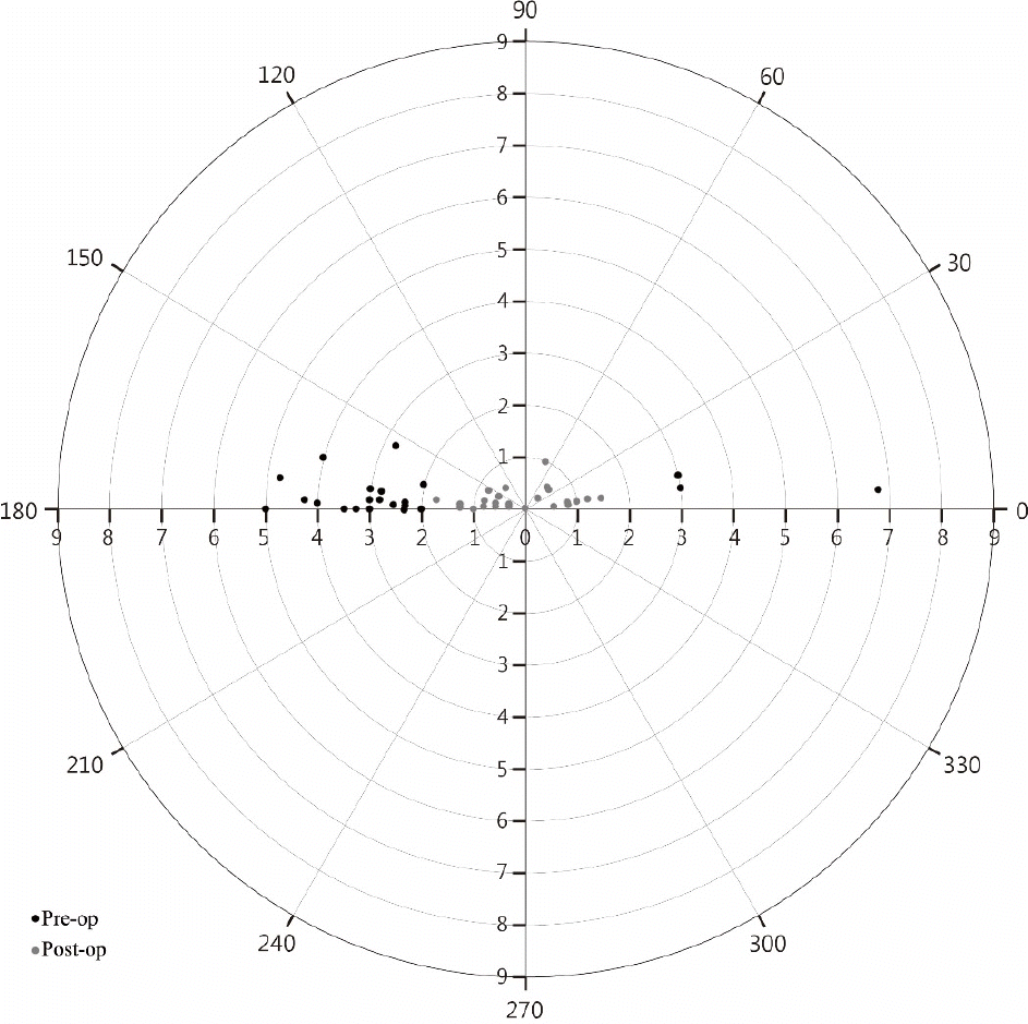
 | Figure 2.Vector analysis of astigmatism. This polar plot shows the reduction in astigmatism after implantable collamer lens im-plantation with simultaneous astigmatic keratotomy. Gray dots (postoperative) are closer to the center compared to black dots (preoperative). Pre-op = preoperation; Post-op = postoperation. |
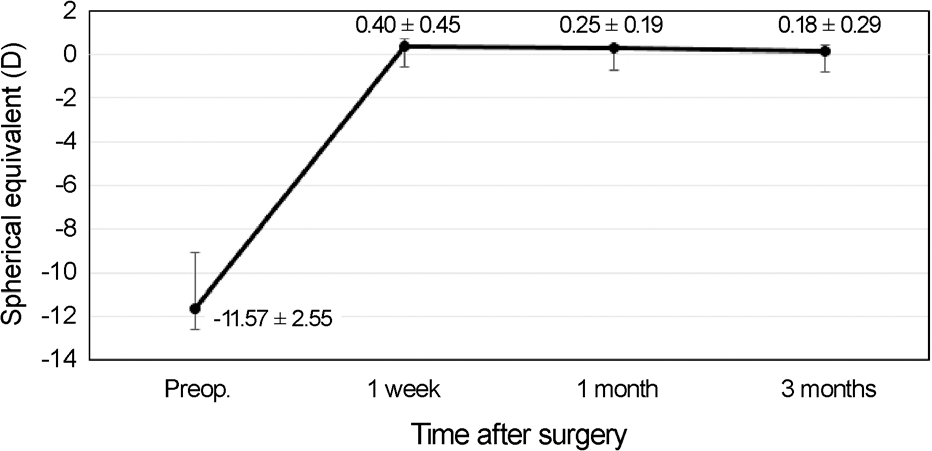
 | Figure 3.Stability of the implantable collamer lens im-plantation with simultaneous astigmatic keratotomy. Spherical equivalent refraction of combined procedure is stable for 3 months. Preop = preoperation. |
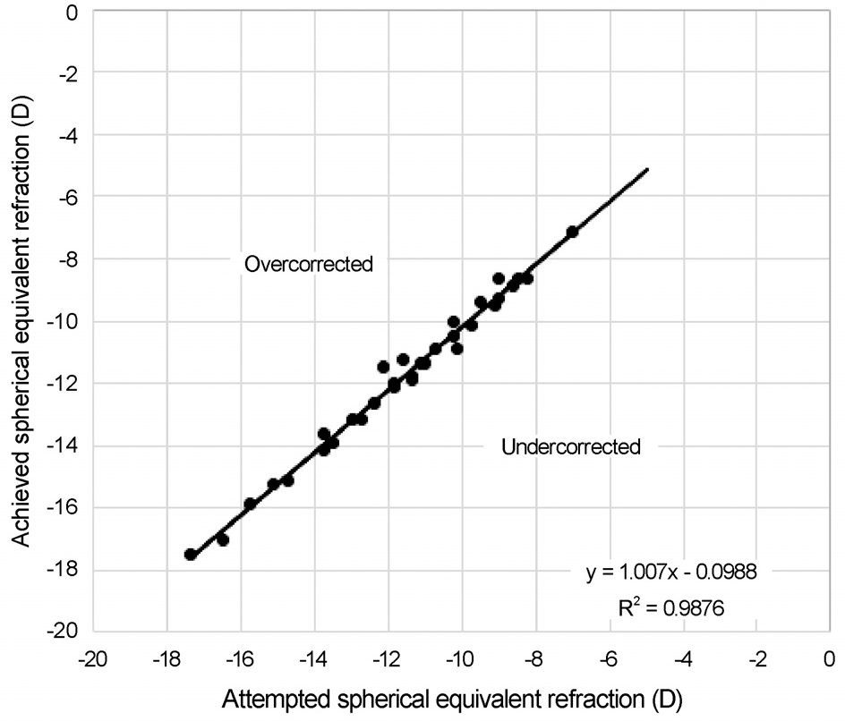
 | Figure 4.Predictability of the implantable collamer lens im-plantation with simultaneous astigmatic keratotomy. Scatterplot of the attempted spherical equivalent refractive change plotted against the achieved spherical equivalent change at 3 months. |
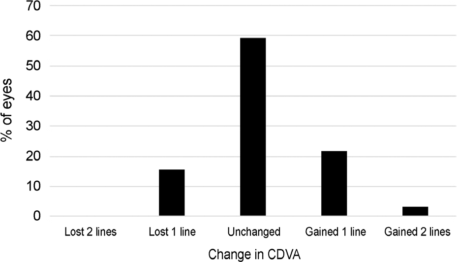
 | Figure 5.Safety of the implantable collamer lens implantation with simultaneous astigmatic keratotomy. The percentage of eyes in which there was a gain/loss of Snellen visual acuity lines (CDVA). CDVA = corrected distance visual acuity. |
Table 1.
Nomogram of the beveled, full-thickness astigmatic ker-atotomy
| Distance from corneal marking (mm) | Incision width (mm) | Corrected astigmatism (D) |
|---|---|---|
| 1.5 | 2.8 | 0.75 |
| 1.5 | 4.1 | 1.25 |
| 1.5 | 5.7 | 2.5 |
| 1.0 | 2.8 | 1 |
| 1.0 | 4.1 | 1.75 |
| 1.0 | 5.7 | 3 |
| 0.5 | 4.1 | 2 |
| 0.5 | 5.7 | 3.5 |
| 0 | 5.7 | 4.5 |
Table 2.
Demographics of the patients
Table 3.
Pre- and post-operative clinical data comparison in patients who underwent implantable collamer lens implantation with si-multaneous astigmatic keratotomy


 PDF
PDF ePub
ePub Citation
 Print
Print


 XML Download