

 PDF
PDF ePub
ePub Citation
 Print
Print


Abstract
Methods
A retrospective chart review of the medical records for 32 eyes of 32 patients who were clinically diagnosed as having a compound nevus from February 2011 to February 2017 was performed.
Results
The average follow-up period was 21.38 (range, 6–70) months for the 32 patients (9 males and 32 females), and the average age was 21 (range, 7–41) years old. The development or detection of a nevus varied between patients. There were no associated symptoms except for one patient who experienced foreign body sensation. An increase in size was noted in 5 cases (15%). The most common location in the conjunctiva was bulbar in 30 cases (93.8%), and the most common quadrant was temporal in 21 cases (65.6%) followed by nasal conjunctiva in 11 cases (34.4%). The most common locations of anterior margin and posterior margin were on the limbus (56%) and bulbar conjunctiva (92%), respectively. The mean horizontal length was 2.59 ± 1.9 mm and the mean vertical length was 2.62 ± 2.1 mm. All horizontal and vertical lengths were within 5 mm. An elevated nevus was observed in 25 cases (78.1%), and 18 cases (56.3%) had cystic lesions. The color of the nevi were largely brown (26 cases, 81.3%), and 29 cases (90.6%) had feeder vessels. Excisional biopsy and histologic exam were performed in 22 cases (68.8%). The purpose of the treatment was mostly cosmetic (20 cases, 93.8%) or for differential diagnosis with malignant melanoma (2 cases, 6.3%). In the surgery group, no one showed recurrence or any significant complications.
Figures and Tables
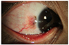
Figure 1
Slit-lamp photographs of compound nevus subtype. Compound nevus which had cystic lesion (A). The color of compound nevus could be pink (B) or yellow (C).
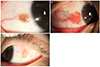
Figure 2
Slit-lamp photographs of compound nevus suspecting malignant. Slit-lamp photograph of a 40-year old female patient shows 4.1 mm (average) diameter sized nevus with dark brown color, irregular margin and feeder vessel (A). Slit-lamp photograph of a 40-years old male patients shows 5.0 mm (average) diameter sized elevated nevus with dark brown color with very irregular margin and multiple feeder vessels (B).
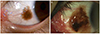
Figure 3
Slit-lamp photographs of compound nevus before excision and 3 months after excision. Compared to preoperative photographs (A, C, E), postoperative photographs (B, D, F) show complete disappearance of pigmentation and good cosmesis.
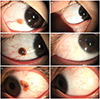
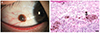
Figure 4
Slit-lamp photograph and histologic feature of compound nevus. (A) Slit-lamp photograph shows pigmented lesion at bulbar conjunctiva. (B) Histologic features of compound nevus (Hematoxylin and eosin stain, ×100) shows that melanocyte forms a nest shape and melanin pigments are in the epidermis (asterisk) and dermis (arrow) (scale bar: 50 µm).
References
1. Folberg R, Jakobiec FA, Bernardino VB, Iwamoto T. Benign conjunctival melanocytic lesions. Clinicopathologic features. Ophthalmology. 1989; 96:436–461.
2. Shields CL, Fasiudden AF, Mashayekhi A, Shields JA. Conjunctival nevi: clinical features and natural course in 410 consecutive patients. Arch Ophthalmol. 2004; 122:167–175.
3. Amoli FA, Heidari AB. Survey of 447 patient with conjunctival neoplastic lesions in Farabi eye hospital, Teheran, Iran. Ophthalmic Epidemiol. 2006; 13:275–279.
4. Yeo HE, Lee SH, Kwon JW. Clinical features of conjunctival nevi in Korean patients. J Korean Ophthalmol Soc. 2009; 50:1510–1513.
5. Jeoung JW, Kim TI, Lee JH, et al. Argon laser ablation of conjunctival nevus. J Korean Ophthalmol Soc. 2004; 45:1989–1994.
6. Shields CL, Regillo AC, Mellen PL, et al. Giant conjunctival nevus: clinical features and natural course in 32 cases. JAMA Ophthalmol. 2013; 131:857–863.
7. Sugiura M, Colby KA, Mihm MC Jr, Zembowicz A. Low-risk and high-risk histologic features in conjunctival primary acquired melanosis with atypia: Clinicopathologic analysis of 29 cases. Am J Surg Pathol. 2007; 31:185–192.
8. Jakobiec FA, Folberg R, Iwamoto T. Clinicopathologic characteristics of premalignant and malignant melanocytic lesions of the conjunctiva. Ophthalmology. 1989; 96:147–166.
9. Yazıcı B, Bilqe AD, Yağcı A, et al. Melanocytic nevus of the tarsal conjunctiva. Balkan Med J. 2016; 33:477–479.
10. Balaeva RN, Kasimov EM. Nevi of conjunctiva as a risk factor of melanoma. Vestn Oftalmol. 2016; 132:21–25.
11. Alsharif AM, Al-Gehedan SM, Alasbali T, et al. Argon laser photoablation for treating benign pigmented conjunctival Nevi. Middle East Afr J Ophthalmol. 2016; 23:247–249.
12. Shin KH, Hwang JH, Kwon JW. Argon laser photoablation of superficial conjunctival nevus: results of 3-year study. Am J Ophthalmol. 2013; 155:823–828.
 XML Download