

 PDF
PDF ePub
ePub Citation
 Print
Print


Abstract
Purpose
To report a case of full-thickness macular hole following intravitreal aflibercept injection in a patient with wet age-related macular degeneration (AMD).
Case summary
A 70-year-old man presented to our department with gradually decreasing vision in his left eye. Best-corrected visual acuity was measured as 0.8 in the right eye and 0.2 in the left eye. Fundus examination, fluorescein angiography, and optical coherence tomography (OCT) showed occult choroidal neovascularization associated with subretinal fluid in the left eye. The patient received several intravitreal ranibizumab and bevacizumab injections in his left eye but responded poorly to the treatment. The patient was switched to intravitreal aflibercept injection. After 1 month, the best corrected visual acuity in the left eye was decreased to 0.05. Although the fundus examination was indistinct, OCT confirmed the presence of a full-thickness macular hole. The patient underwent pars plana vitrectomy with internal limiting membrane peeling and fluid-gas exchange, 20% SF6 gas injection, phacoemulsification, and posterior chamber intraocular lens implantation. One month after the operation, the best corrected visual acuity was 0.2. The macular hole was closed completely, as confirmed by OCT.
Figures and Tables
Figure 1
Fundus photography, fluorescein aniography (FA) and Optical coherence tomography (OCT). (A) Baseline fundus examination shows serous retinal detachment and some drusen. (B) Late phase FA shows occult choroidal neovascularization (CNV). (C) Baseline OCT shows type 1 CNV with subretinal fluid. (D) After treatment with 14 times intravitreal ranibizumab injections, OCT showed complete absorption of subretinal fluid. (E) This patient was treated with three intravitreal bevacizumab injections. OCT showed intraretinal fluid, increased retinal pigment epithelial detachment (RPED) and CNV. (F) Two weeks after the first intravitreal aflibercept injection, OCT showed a posterior viterous detachment, subretinal fluid, decrease of RPED and impending macular hole.
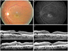
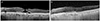
Figure 2
Optical coherence tomography (OCT). (A) One month after intravitreal aflibercept injection, OCT showed a full thickness macular hole. (B) One month after the vitrectomy, the internal limiting membrane peeling and the fluid-gas exchange, OCT showed that the macular hole was closed completely.
References
1. Heier JS, Brown DM, Chong V, et al. Intravitreal aflibercept (VEGF trap-eye) in wet age-related macular degeneration. Ophthalmology. 2012; 119:2537–2548.
2. Jo YJ, Kim KN, Lee JE, Kim JY. Macular hole following intravitreal ranibizumab injections for choroidal neovascularization. J Korean Ophthalmol Soc. 2010; 51:774–778.
3. Kim JM, Jang JW, Kyung SE, Chang MH. Macular hole after single intravitreal injection of ranibizumab in a patient with age-related macular degeneration. J Korean Ophthalmol Soc. 2013; 54:1130–1134.
4. Oshima Y, Apte RS, Nakao S, et al. Full thickness macular hole case after intravitreal aflibercept treatment. BMC Ophthalmol. 2015; 15:30.
5. Kim JH, Cho NC, Kim WJ. Intravitreal aflibercept for neovascular age-related macular degeneration resistant to bevacizumab and ranibizumab. J Korean Ophthalmol Soc. 2015; 56:1359–1364.
6. Fung AE, Rosenfeld PJ, Reichel E. The International Intravitreal Bevacizumab Safety Survey: using the internet to assess drug safety worldwide. Br J Ophthalmol. 2006; 90:1344–1349.
7. Shima C, Sakaguchi H, Gomi F, et al. Complications in patients after intravitreal injection of bevacizumab. Acta Ophthalmol. 2008; 86:372–376.
8. Moisseiev E, Goldstein M, Loewenstein A, Moisseiev J. Macular hole following intravitreal bevacizumab injection in choroidal neovascularization caused by age-related macular degeneration. Case Rep Ophthalmol. 2010; 1:36–41.
9. Okamoto T, Shinoda H, Kurihara T, et al. Intraoperative and fluorescein angiographic findings of a secondary macular hole associated with age-related macular degeneration treated by pars plana vitrectomy. BMC Ophthalmol. 2014; 14:114.
10. Raiji VR, Eliott D, Sadda SR. Macular hole overlying pigment epithelial detachment after intravitreal injection with ranibizumab. Retin Cases Brief Rep. 2013; 7:91–94.
 XML Download