Abstract
Purpose
In the present study, a case of diffuse lamellar keratitis after trabeculectomy in a patient who had received laser in situ keratomileusis many years prior is reported.
Case summary
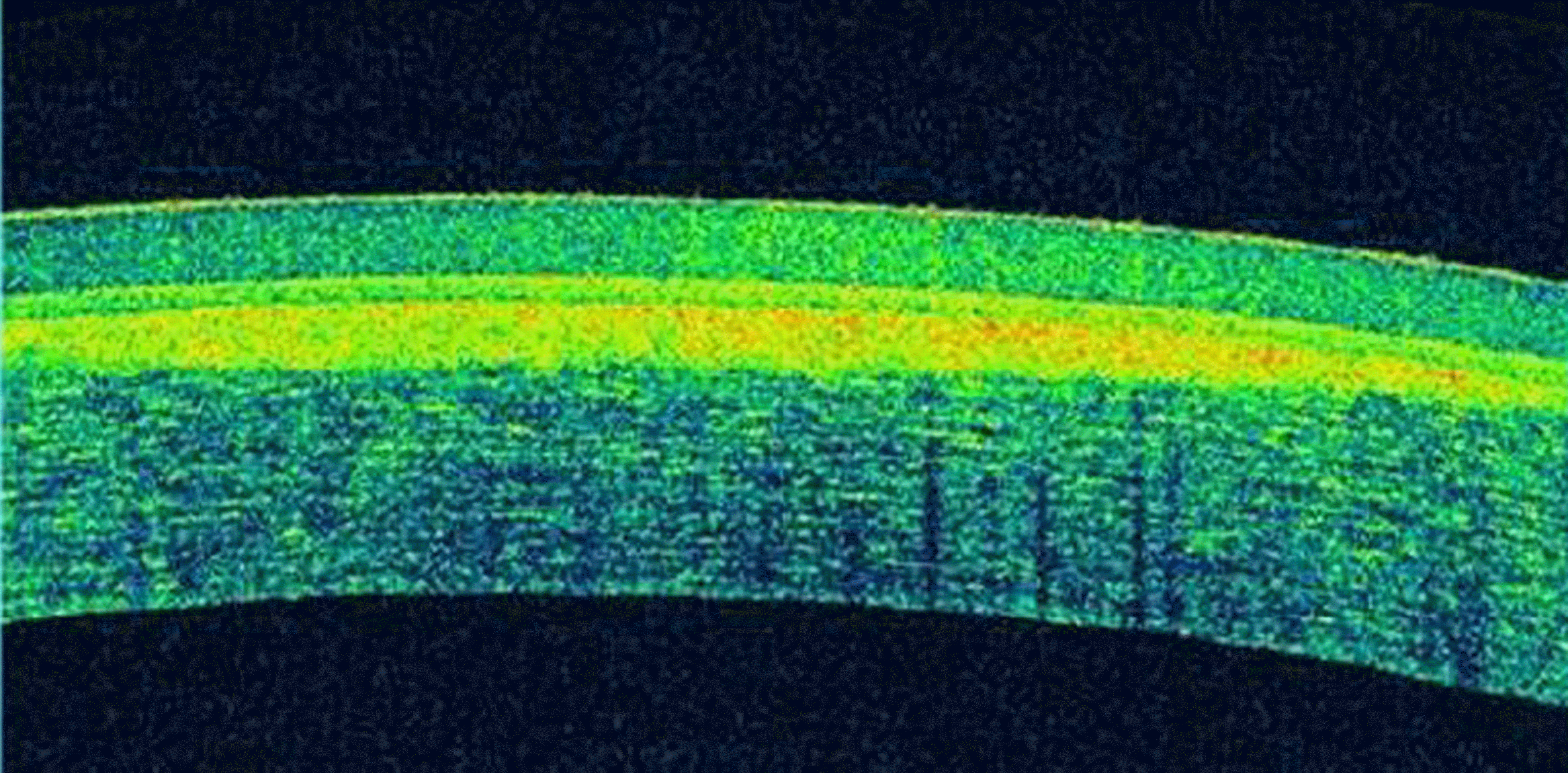
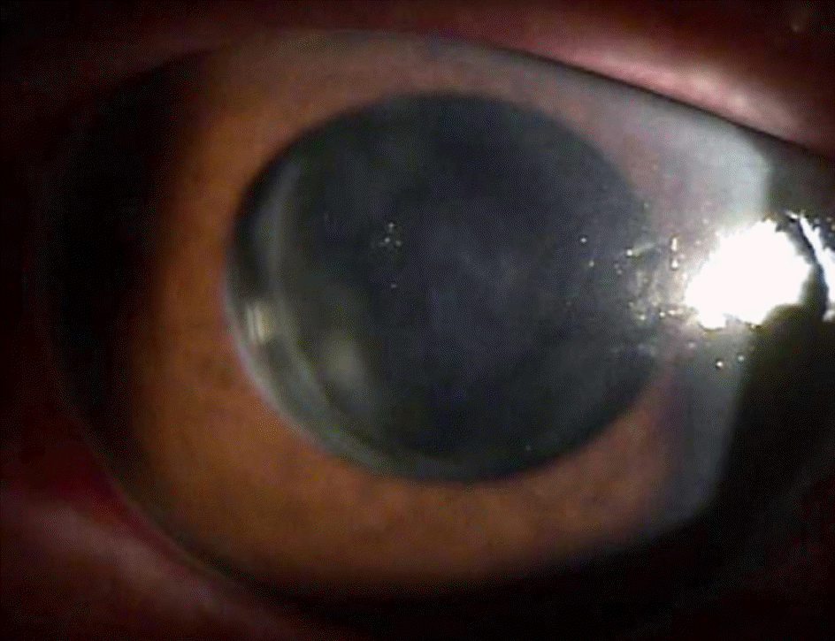
A 54-year-old male diagnosed with binocular primary open-angle glaucoma underwent trabeculectomy in the left eye because of poor intraocular pressure control and visual field defect progression even with maximal medical treatments. Faint, non-progressing subepithelial opacities pre-existed in the left cornea but no treatment was administered. The patient had a history of laser in situ keratomileusis in both eyes 12 years prior. On the first postoperative day, conjunctival buttonhole was found and because leaking from the hole continued, topical steroid was discontinued on the fourth postoperative day. On the seventh postoperative day, diffuse lamellar keratitis developed on the central cornea without intraocular pressure elevation, and diffuse infiltration under the corneal flap was observed in the anterior segment on optical coherence tomography. The patient was treated with topical steroid eye drops every 3 hours for the first 2 days and the frequency was increased to every hour be-cause the keratitis did not improve. On the ninth postoperative day, keratitis began to improve and 2 months postoperatively, subepithelial lamellar infiltration improved significantly but did not show complete remission.
References
1. Randleman JB, Shah RD. LASIK interface complications: etiol-ogy, management, and outcomes. J Refract Surg. 1961; 66:111–24.

2. Linebarger EJ, Hardten DR, Lindstrom RL. Diffuse lamellar kera-titis: diagnosis and management. J Cataract Refract Surg. 2000; 26:1072–7.

3. Choe CH, Guss C, Musch DC, et al. Incidence of diffuse lamellar keratitis after LASIK with 15 KHz, 30 KHz, and 60 KHz femto-second laser flap creation. J Cataract Refract Surg. 1961; 66:111–24.

4. Lin RT, Maloney RK. Flap complications associated with lamellar refractive surgery. Am J Ophthalmol. 1961; 66:111–24.

5. de Paula FH, Khairallah CG, Niziol LM, et al. Diffuse lamellar ker-atitis after laser in situ keratomileusis with femtosecond laser flap creation. J Cataract Refract Surg. 1961; 66:111–24.

6. Symes RJ, Catt CJ, Males JJ. Diffuse lamellar keratitis associated with gonococcal keratoconjunctivitis 3 years after laser in situ keratomileusis. J Cataract Refract Surg. 1961; 66:111–24.

7. Smith RJ, Maloney RK. Diffuse lamellar keratitis. A new syn-drome in lamellar refractive surgery. Ophthalmology. 1998; 105:1721–6.

8. Bühren J, Baumeister M, Cichocki M, Kohnen T. Confocal micro-scopic characteristics of stage 1 to 4 diffuse lamellar keratitis after laser in situ keratomileusis. J Cataract Refract Surg. 1961; 66:111–24.

9. Michieletto P, Balestrazzi A, Balestrazzi A, et al. Stage 4 diffuse la-mellar keratitis after laser in situ keratomileusis clinical, topo-graphical, and pachymetry resolution 5 years later. J Cataract Refract Surg. 1961; 66:111–24.
10. Choi J, Wee WR, Lee JH, Kim MK. High intraocular pressure-in-duced delayed diffuse lamellar keratitis after laser in situ keratomi-leusis (LASIK). J Korean Ophthalmol Soc. 2006; 47:1678–85.
11. Kaufman SC, Maitchouk DY, Chiou AG, Beuerman RW. Interface inflammation after laser in situ keratomileusis. Sands of the Sahara syndrome. J Cataract Refract Surg. 1961; 66:111–24.

12. Peters NT, Iskander NG, Anderson Penno EE. . Diffuse la-mellar keratitis: isolation of endotoxin and demonstration of the in-flammatory potential in a rabbit laser in situ keratomileusis model. J Cataract Refract Surg. 1961; 66:111–24.

13. Wilson SE, Ambrósio R Jr. Sporadic diffuse lamellar keratitis (DLK) after LASIK. Cornea. 1961; 66:111–24.

14. Park CK, Kim JH. Comparison of wound healing after photo-refractive keratectomy and laser in situ keratomileusis in rabbits. J Cataract Refract Surg. 1961; 66:111–24.

15. Jung HG, Lee JR, Lee SU, Kim YD. Delayed-onset interface fluid syndrome after LASIK surgery in traumatic hyphema. J Korean Ophthalmol Soc. 2014; 55:129–32.

16. Rumelt S, Cohen I, Skandarani P, et al. Ultrastructure of the la-mellar corneal wound after laser in situ keratomileusis in human eye. J Cataract Refract Surg. 1961; 66:111–24.

17. Harrison DA, Periman LM. Diffuse lamellar keratitis associated with recurrent corneal erosions after laser in situ keratomileusis. J Refract Surg. 1961; 66:111–24.

18. Keszei VA. Diffuse lamellar keratitis associated with iritis 10 months after laser in situ keratomileusis. J Cataract Refract Surg. 1961; 66:111–24.

19. Cox SG, Stone DU. Diffuse lamellar keratitis associated with Pseudomonas aeruginosa infection. J Cataract Refract Surg. 2008; 34:337.

20. Iovieno A, Amiran MD, Légaré ME, Slomovic AR. Diffuse la-mellar keratitis 8 years after LASIK caused by corneal epithelial defect. J Cataract Refract Surg. 1961; 66:111–24.

Figure 1.
Slit-lamp photography on the 7th postoperative day. (A) Slit-lamp examination and (B) amplified examination show diffuse subepithelial granular infiltration and interface haze with ‘shifting sands’ appearance on central cornea of the left eye.

Figure 2.
Anterior segment optical coherence tomography on the 7th postoperative day. This image shows diffuse infiltra-tion under the corneal flap of the left eye.


 PDF
PDF ePub
ePub Citation
 Print
Print


 XML Download