Abstract
Purpose
In the present study, a case of IgG4-related ophthalmic disease which met the 2015 IgG4-related ophthalmic disease diagnostic criteria is reported and literature review performed.
Case summary
A 51-year-old female presented with both upper eyelid swelling, redness, and a palpable mass. Eye movements were normal and exophthalmos was not observed. Facial computed tomography showed both lacrimal gland hypertrophy. The patient underwent left anterior orbitotomy with incisional biopsy. Immunostained biopsy showed the ratio of IgG4+ to IgG+ cells was 50% and the mean number of IgG4-positive plasma cells was approximately 150 per high-power field. Hematological examination showed elevated serum IgG4 concentrations of 6,930 mg/dL, however, other organs were not involved. The patient satisfied the diagnostic criteria for IgG4-related ophthalmic disease. The patient was given an oral steroid and immunosuppressant and the symptoms improved.
Figures and Tables
 | Figure 1Photograph and orbital computed tomography of the patient. (A) Both upper eyelid swelling was observed at the first visit. (B) Orbital computed tomography revealed bilaterally enlarged lacrimal glands. |
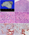
 | Figure 2Biopsied specimens from lacrimal gland. (A) Gross finding of the eyelid mass. (B) Low-power view of the mass showed diffuse lymphoid proliferation consisting of hyperplastic follicles of variable size and intervening sclerotic area (H-E stain, ×20). (C, D) High-power view of the mass showed entrapped epithelial glands in the sclerotic stroma and a lot of plasma cells (arrows) (H-E stain, ×400). (E) There were many plasma cells with IgG immunoreactivity (IgG, ×100, Inset: ×400). The mean number of IgG-positive plasma cells (arrows) was about 300 per a high-power field. (F) There were many plasma cells with IgG4 immunoreactivity (IgG4, ×100, Inset: ×400). The mean number of IgG4-positive plasma cells (arrows) was about 150 per a high-power field. |
References
1. Umehara H, Okazaki K, Masaki Y, et al. Comprehensive diagnostic criteria for IgG4-related disease (IgG4-RD), 2011. Mod Rheumatol. 2012; 22:21–30.
2. Goto H, Takahira M, Azumi A. Japanese Study Group for IgG4-Related Ophthalmic Disease. Diagnostic criteria for IgG4-related ophthalmic disease. Jpn J Ophthalmol. 2015; 59:1–7.
3. Kim K, Lee MJ, Kim NJ, et al. Three cases of Hyper-IgG4 syndrome involving ocular adnexa. J Korean Ophthalmol Soc. 2010; 51:1133–1138.
4. Cho HS, Choi JY, Yum JH. A case of IgG4-related sclerosing disease involving the optic nerve. J Korean Ophthalmol Soc. 2012; 53:1879–1884.
5. Yoon JH, Jung JW, Chi MJ. A case of IgG4-related sclerosing disease involving the eyelid in an idiopathic sclerosing myositis patient. J Korean Ophthalmol Soc. 2013; 54:160–164.
6. Wallace ZS, Deshpande V, Stone JH. Ophthalmic manifestations of IgG4-related disease: single-center experience and literature review. Semin Arthritis Rheum. 2014; 43:806–817.
7. Kubota T, Katayama M, Moritani S, Yoshino T. Serologic factors in early relapse of IgG4-related orbital inflammation after steroid treatment. Am J Ophthalmol. 2013; 155:373–379.


 PDF
PDF ePub
ePub Citation
 Print
Print


 XML Download