

 PDF
PDF ePub
ePub Citation
 Print
Print


Abstract
Purpose
To evaluate the efficacy of optical coherence tomography angiography (OCTA) by measuring the foveal avascular zone (FAZ) area in patients with branch retinal vein occlusion (BRVO).
Methods
Thirty four eyes of 34 patients with BRVO were retrospectively reviewed. The area of the FAZ was calculated using flu-orescein angiography (FAG) and OCTA. The FAZ area was divided into two groups according to the presence of macular ede-ma, which was determined based on the central foveal thickness (300 μ m), and then the measured areas were compared.
Results
Pearson’s correlation analysis revealed a significant positive correlation between FAG and the superficial capillary plexus (SCP) in OCTA with or without macular edema (r = 0.845, p = 0.001). However, there was not a significant correlation be-tween FAG and the deep capillary plexus (DCP) in OCTA (r = 0.001, p = 0.996). In addition, the FAZ area measured by FAG and OCTA in the SCP showed a significant agreement between the two methods (intraclass correlation coefficient [ICC] = 0.916, p = 0.001). However, there was no significant relation found for the FAZ area between FAG and OCTA in the DCP (ICC = 0.001, p = 0.501).
Conclusions
In the patients with BRVO, OCTA can be used to measure the FAZ areas in both the SCP and DCP, beyond meas-urement of the FAZ area at the two-dimensional cross section used during FAG. The FAZ area in the SCP via OCTA showed a statistically significant correlation with the FAZ area determined by FAG, but there was no such correlation in the DCP. That said, the FAZ area in the DCP was positively correlated with a decrease in visual acuity among the patients, which may be an indicator of visual prognosis.
Go to : 
References
1. Orth DH, Patz A. Retinal branch vein occlusion. Surv Ophthalmol. 1978; 22:357–76.

2. Chung JH, Choi GJ, Na KS. The macular circulation state on BRVO according to occlusion site. J Korean Ophthalmol Soc. 2000; 41:1556–62.
3. Kang SJ, Chin HS, Moon YS. Visual prognosis of macular edema associated with macular ischemia in branch retinal vein occlusion. J Korean Ophthalmol Soc. 2002; 43:1621–8.
4. Parodi MB, Visintin F, Della Rupe P, Ravalico G. Foveal avascular zone in macular branch retinal vein occlusion. Int Ophthalmol. 1995; 19:25–8.
5. Wu L. Treatments for retinal vein occlusion: a review of recent developments. Retina Today. 2014; 4:62–3.
6. Brown DM. Clinical implications of the BRAVO and CRUISE trials. How should physicians apply this new information in their treatment of CRVO and BRVO. Retina Today. 2010; 5:38–40.
7. Yannuzzi LA, Rohrer KT, Tindel Lj. . Fluorescein angiography complication survey. Ophthalmology. 1986; 93:611–7.
8. Staurenghi G, Invernizzi A, de Polo L, Pellegrini M. Macular edema. Diagnosis and detection. Dev Ophthalmol. 2010; 47:27–48.
9. Shrout PE, Fleiss JL. Intraclass correlations: uses in assessing rater reliability. Psychol Bull. 1979; 86:420–8.
10. Rogers SL, McIntosh RL, Lim L. . Natural history of branch retinal vein occlusion: an evidence-based systematic review. Ophthalmology. 2010; 117:1094–101.e5..
11. Fong DS, Ferris FL, Davis MD. . Causes of severe visual loss in the early treatment diabetic retinopathy study: ETDRS report no. 24. Early Treatment Diabetic Retinopathy Study Research Group. Am J Ophthalmol. 1999; 127:137–41.
12. Bresnick GH, Condit R, Syrjala S. . Abnormalities of the fo-veal avascular zone in diabetic retinopathy. Arch Ophthalmol. 1984; 102:1286–93.
13. Arend O, Wolf S, Harris A, Reim M. The relationship of macular microcirculation to visual acuity in diabetic patients. Arch Ophthalmol. 1995; 113:610–4.
14. Mansour AM. Measuring fundus landmarks. Invest Ophthalmol Vis Sci. 1990; 31:41–2.
15. Ryan SJ, editor. Retina, 4th ed. Los Angeles: Elsevier;2006. p. 541.
16. Argon laser photocoagulation for macular edema in branch vein occlusion. The Branch vein occlusion Study group. Am J Ophtahalmol. 1984; 98:271–82.
17. Arend O, Wolf S, Jung F. . Retinal microcirculation in patients with diabetes mellitus: dynamic and morphological analysis of perifoveal capillary network. Br J Ophthalmol. 1991; 75:514–8.
18. Lee SJ, Koh HJ. Enlargement of the foveal avascular zone in dia-betic retinopathy after adjunctive intravitreal bevacizumab (avastin) with pars plana vitrectomy. J Ocul Pharmacol Ther. 2009; 25:173–4.
19. Stein MR, Parker CW. Reactions following intravenous fluorescein. Am J Ophthalmol. 1971; 72:861–8.
20. Karhunen U, Raitta C, Kala R. Adverse reactions to fluorescein angiography. Acta Ophthalmol (Copenh). 1986; 282–6.
21. Lipson BK, Yannuzzi LA. Coplication of intravenous fluorescein injections. Int Ophthalmol Clin. 1989; 29:200–5.
22. Amalric P, Biau C, Féniès MT. Incidents and accidents during fluo-rescein angiography. Bull Soc Ophthalmol Fr. 1968; 68:968–73.
23. Deglin SM, Deglin EA, Chung EK. Acute mycardial infacrtion fol-lowing fluorescein angiography. Heart Lung. 1977; 6:505–9.
24. Cunnigham EE, Balu V. Cardiac arrest following fluorescein angiography. JAMA. 1979; 242:2431.
25. Wons J, Pfau M, Wirth MA. . Optical coherence tomography angiography of the foveal avascular zone in retinal vein occlusion. Ophtalmologica. 2016; 235:195–202.
26. Martinet V, Guigui B, Glacet-Bernard A. . Macular edema in central retinal vein occlusion: correlation between optical coher-ence tomography, angiography and visual acuity. Int Ophthalmol. 2012; 32:369–77.
27. Ghasemi Falavarjani K, Iafe NA, Hubschman JP. . Optical co-herence tomography angiography analysis of the foveal avascular zone and macular vessel density after anti-VEGF therapy in eyes with diabetic macular edema and retinal vein occlusion. Invest Ophthalmol Vis Sci. 2017; 58:30–4.
28. Lee J, Moon BG, Cho AR, Yoon YH. Optical coherence tomog-raphy angiography of DME and its association with anti-VEGF treatment response. Ophthalmology. 2016; 123:2368–75.
Go to :
 | Figure 1.Branch retinal vein occlusion without macular edema. Color fundus photo (A), Fluorescein angiography (FAG; B), Zoom on the fovea in FAG (C), optical coherence tomography angiography (OCTA) in the superficial capillary plexus showed quite nor-mal and well demarcated foveal capillary arcade (D). OCTA in the deep capillary plexus showed dilatation of deep inferior macular capillaries with hyper signal although there was not significant magnified nonperfused area in fovea (E). Optical coherence tomog-raphy showed there was no intraretinal cystic lesion (F). |
 | Figure 2.Macular edema related to a branch retinal vein occlusion. Color fundus photo (A), Fluorescein angiography (FAG; B), Zoom on the fovea in FAG (C), optical coherence tomography angiography (OCTA) in the superficial capillary plexus showed pret-ty well demarcated foveal capillary arcade although there was disruption of the capillary network superiorly (D). However, OCTA in the deep capillary plexus (DCP) showed central black cysts, disorganization of the deep superior macula and magnified non-perfused area (E). Optical coherence tomography showed multiple intraretinal cysts where the corresponding OCTA in the DCP (F). |
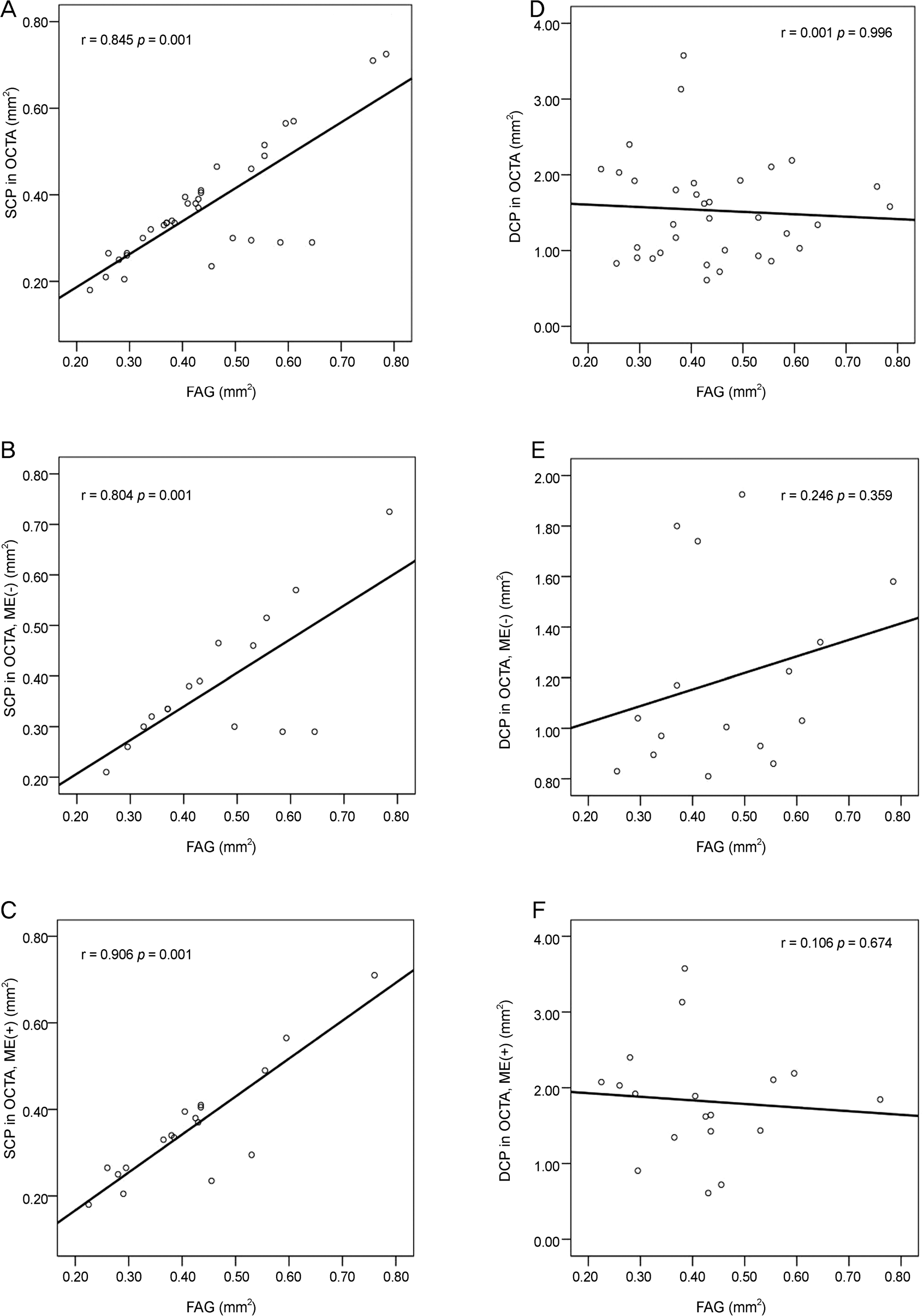 | Figure 3.Relationship of foveal avascular zone area measured between fluorescein angiography (FAG) and optical coherence to-mography angiography (OCTA). Pearson’s correlation analysis showed significant positive correlation between FAG and superficial capillary plexus (SCP) in OCTA with or without macular edema (ME) (A: FAG vs. SCP in OCTA, r = 0.845, p = 0.001; B: FAG vs. SCP in OCTA without ME, r = 0.804, p = 0.001; C: FAG vs. SCP in OCTA with ME, r = 0.906, p = 0.001). But, there was not significant correlation between FAG and deep capillary plexus (DCP) in OCTA (D: FAG vs. DCP in OCTA, r = 0.001, p = 0.996; E: FAG vs. DCP in OCTA without ME, r = 0.246, p = 0.359; F: FAG vs. DCP in OCTA with ME, r = 0.106, p = 0.674). |
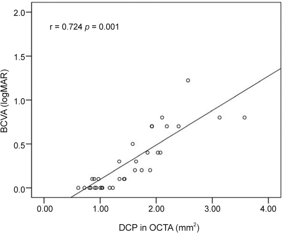 | Figure 4.Relationship of best corrected visual acuity (BCVA; logMAR) and deep capillary plexus (DCP) in optical coherence tomography angiography (OCTA). Pearson's correlation analysis showed significant positive correlation between logMAR and DCP in OCTA (r = 0.724 p = 0.001). logMAR = logarithm of the minimum angle of resolution. |
Table 1.
Subject baseline demographic and ocular characteristics
| Characteristics | Total subjects | Macular edema | p-value* | |
|---|---|---|---|---|
| (n = 34) | (-) (n = 16) | (+) (n = 18) | ||
| Age (years) | 60.94 ± 9.75 | 60.56 ± 9.44 | 61.28 ± 10.27 | 0.835 |
| Male/Female | 15/19 | 7/9 | 8/10 | 1.000 |
| BCVA (logMAR) | 0.27 ± 0.35 | 0.22 ± 0.42 | 0.32 ± 0.28 | 0.044 |
| HTN | 16 | 7 | 9 | 0.984 |
| DM | 5 | 1 | 4 | 0.408 |
| Hyperlipidemia | 4 | 2 | 2 | 1.000 |
| Central foveal thickness (μ m) | 327.12 ± 120.98 | 246.38 ± 31.98 | 398.89 ± 122.38 | 0.001 |
Table 2.
The intraclass correlation coefficient (ICC) was used to determine the interobserver reproducibility for the manually meas ured foveal avascular zone (FAZ)
Table 3.
Comparisons of foveal avascular zone (FAZ) areas with fluorescein angiography (FAG) and optical coherence tomography angiography (OCTA) in branch retinal vein occlusion with or without macular edema and normal fellow eyes
| FAZ area (mm2) | Normal fellow eyes | Macular edema | p-value | |
|---|---|---|---|---|
| (-) (n = 16) | (+) (n = 18) | p-value | ||
| FAG | 0.46 ± 0.14 | 0.46 ± 0.12 | 0.863* | |
| SCP in OCTA | 0.39 ± 0.13 | 0.38 ± 0.13 | 0.36 ± 0.13 | 0.547† |
| DCP in OCTA | 1.09 ± 0.23 | 1.20 ± 0.37 | 1.83 ± 0.75 | 0.001† |
Table 4.
Comparison of intraclass correlations of foveal avascular zone (FAZ) area measurements from fluorescein angiography (FAG) and optical coherence tomography angiography (OCTA) in branch retinal vein occlusion patients
 XML Download