

 PDF
PDF ePub
ePub Citation
 Print
Print


Abstract
Purpose
Choristomas represent congenital overgrowth of normal tissues in an abnormal location. The simultaneous presence of epibulbar choristoma and microphthalmos has rarely been reported. The authors report a case of extensive epibulbar chori-stoma associated with microphthalmos.
Case summary
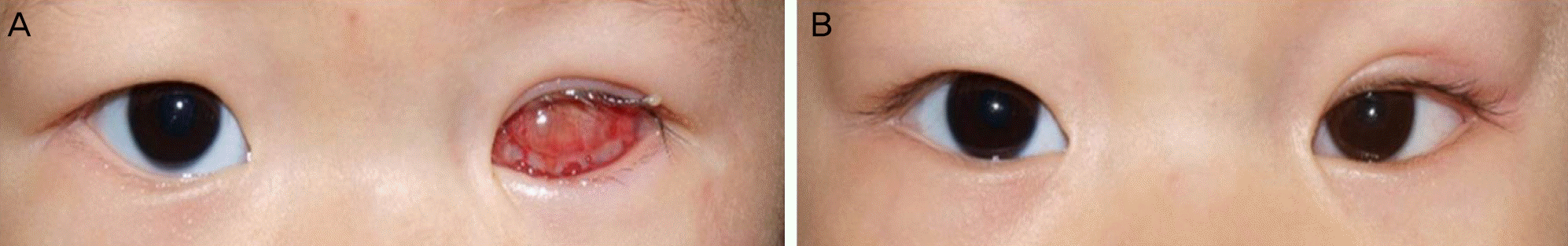
A 9-day-old boy with the left eyeball absent from birth was referred to our clinic. A large cornea-like structure covered by keratinized membrane was observed inside the eyelid aperture, therefore buphthalmos or corneal staphyloma with microphthalmos was presumed. At the age of 2 months, a large mass of central conjunctival sac protruded through the left eyelid aperture. Manual reduction could not return the tissue to its original site and the mass immediately protruded again. At the age of 9 months, orbital magnetic resonance imaging showed the small presumed ocular tissue behind the large mass of fat signal in the central anterior orbit, therefore, extensive epibulbar choristoma associated with microphthalmos was diagnosed. At 12 months of age, partial excision of the protruding portion of the mass was performed. Based on pathologic examination, the mass was determined to be a choristoma and cosmetically acceptable appearance with prosthesis was maintained for 10 months after the surgery.
References
1. Mansour AM, Barber JC, Reinecke RD, Wang FM. Ocular choristomas. Surv Ophthalmol. 1961; 66:111–24.

2. Hou Z, Korn BS, Ding J, Li D. Management of extensive epibulbar choristoma associated with microphthalmos: a rare clinical entity. JAMA Ophthalmol. 1961; 66:111–24.
3. Schulze RR. Limbal dermoid tumor with intraocular extension. Arch Ophthalmol. 1961; 66:111–24.
4. Chawla B, Chauhan K, Kashyap S. Mature orbital teratoma with an ectopic tooth and primary anophthalmos. Orbit. 1961; 66:111–24.
5. Casey RJ, Garner A. Epibulbar choristoma and microphthalmia: a report of two cases. Br J Ophthalmol. 1961; 66:111–24.
6. Murata T, Ishibashi T, Ohnishi Y, Inomata H. Corneal choristoma with microphthalmos. Arch Ophthalmol. 1961; 66:111–24.
7. Huang TY, Tsai YJ, Tan HY, Ma L. Managing epibulbar chori-stoma with microphthalmos. J Pediatr Ophthalmol Strabismus. 1961; 66:111–24.
Figure 1.
The photographs and orbital magnetic resonance imaging (MRI) of the patient. (A) At the age of two months, large mass of central conjunctival sac protruded through the eyelid aperture of the left eye. (B) Orbital MRI at the age of 9 months showed the small presumed ocular tissue behind the large fat-signaled mass with focal cyst in central anterior orbit. (C, D) Partial excision of protruding mass from conjunctival sac was done at 12 months of age.
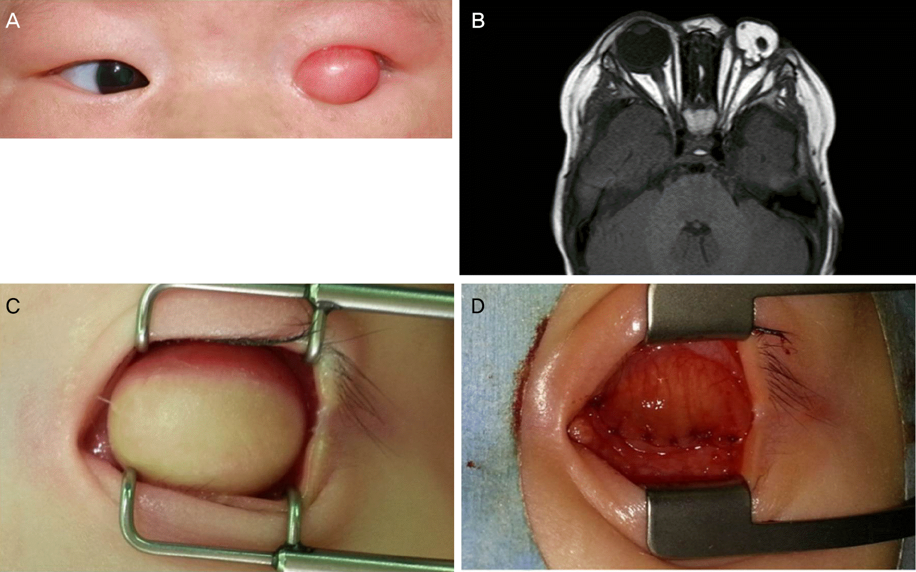
Figure 2.
The photographs of histopathological findings. (A, B) The low magnified images (A: Hematoxylin and eosin [HE] stain, 12.5 magnification, B: HE stain, ×40 magnification): the lesion was covered anteriorly by mature, keratinized stratified squamous epithelium and dermis. Deep to the dermis was adipose tissue. (C) The high magnified image (HE stain, ×100 magnification): the lesion exhibits patches of focal lacrimal ducts (arrow), dilated apocrine duct (arrowhead) and areas of adipose tissue (*).

 XML Download