

 PDF
PDF ePub
ePub Citation
 Print
Print


Abstract
Purpose
To analyze the relationship between ocular surface disease index and tear film lipid layer thickness (LLT) using a LipiView II® (LipiView® Ocular Surface Interferometer, TearScience®, Morrisville, NC, USA) interferometer.
Methods
Forty-nine patients diagnosed with dry eye syndrome were recruited for this prospective study. Patients completed oc-ular surface disease index questionnaires. We performed slit lamp examination, Schirmer test, corneal and conjunctival fluo-rescein staining, measured tear film break-up time, and graded meibomian gland dysfunction. Tear LLT, blinking time, and dy-namic meibomian imaging were analyzed using a LipiView II® ocular interferometer. To control for missing data, we analyzed four sets of imputated data via the multiple imputation method and performed Pearson correlation analysis. Patients were as-signed to one of two LLT categories (LLT < 60 or LLT ≥ 60) and Chi-square test was performed.
Results
Among ocular surface disease parameters, tear film break-up time (tBUT) had a statistically significant correlation with average and maximum LLT (average LLT; p = 0.008, 0.035, 0.006, 0.049, maximum LLT; p = 0.006, 0.042, 0.020, 0.049, Pearson correlation analysis with multiple imputation) but there was no significant correlation with minimum LLT (minimum LLT; p = 0.048, 0.090, 0.079, 0.039). Of the patients with a relatively thick average LLT or maximum LLT (LLT ≥ 60 nm), 80% and 88% had a tBUT < 10, respectively. Conversely, 39% and 47% of patients with relatively thin average LLT (LLT < 60 nm) had a tBUT < 10 (average LLT; p = 0.013, maximum LLT; p = 0.039).
Conclusions
Average LLT and maximum LLT were significantly correlated with tBUT. Patients with a relatively thin average or maximum LLT tended to have a shorter tBUT. Based on these results, measuring tear film LLT using a LipiView II® interferometer may be useful in the diagnosis and follow-up of patients with evaporative dry eye.
Go to : 
References
1. Shine WE, McCulley JP. The role of cholesterol in chronic blepharitis. Invest Ophthalmol Vis Sci. 1991; 32:2272–80.
2. Shine WE, McCulley JP. Role of wax ester fatty alcohols in chronic blepharitis. Invest Ophthalmol Vis Sci. 1993; 34:3515–21.
3. Shine WE, McCulley JP. Keratoconjunctivitis sicca associated with meibomian secretion polar lipid abnormality. Arch Ophthalmol. 1998; 116:849–52.

4. Shine WE, McCulley JP. Polar lipids in human meibomian gland secretions. Curr Eye Res. 2003; 26:89–94.
5. Bron AJ, Tiffany JM. The contribution of meibomian disease to dry eye. Ocul Surf. 2004; 2:149–65.
6. Foulks GN, Bron AJ. Meibomian gland dysfunction: a clinical scheme for description, diagnosis, classification, and grading. Ocul Surf. 2003; 1:107–26.
7. Geerling G, Tauber J, Baudouin C. . The international work-shop on meibomian gland dysfunction: report of the subcommittee on management and treatment of meibomian gland dysfunction. Invest Ophthalmol Vis Sci. 2011; 52:2050–64.
8. Tomlinson A, Bron AJ, Korb DR. . The international workshop on meibomian gland dysfunction: report of the diagnosis sub- committee. Invest Ophthalmol Vis Sci. 2011; 52:2006–49.
9. Greiner JV, Glonek T, Korb DR. . Volume of the human and rab-bit meibomian gland system. Adv Exp Med Biol. 1998; 438:339–43.
10. Chew CK, Jansweijer C, Tiffany JM. . An instrument for quan-tifying meibomian lipid on the lid margin: the Meibometer. Curr Eye Res. 1993; 12:247–54.
11. Blackie CA, Solomon JD, Scaffidi RC. . The relationship be-tween dry eye symptoms and lipid layer thickness. Cornea. 2009; 28:789–94.
12. Finis D, Pischel N, Schrader S, Geerling G. Evaluation of lipid lay-er thickness measurement of the tear film as a diagnostic tool for Meibomian gland dysfunction. Cornea. 2013; 32:1549–53.
13. The definition and classification of dry eye disease: report of the Definition and Classification Subcommittee of the International Dry Eye WorkShop (2007). Ocul Surf. 2007; 5:75–92.
14. Jeon SJ, Baek JW, Doh SH, Chung SK. Tear meniscus evaluation using optical coherence tomography in meibomein gland dysfunction patients. J Korean Ophthalmol Soc. 2015; 56:1684–91.
15. Arita R, Itoh K, Inoue K, Amano S. Noncontact infrared meibog-raphy to document age-related changes of the meibomian glands in a normal population. Ophthalmology. 2008; 115:911–5.
16. Mathers WD. Ocular evaporation in meibomian gland dysfunction and dry eye. Ophthalmology. 1993; 100:347–51.
17. Mathers WD, Lane JA. Meibomian gland lipids, evaporation, and tear film stability. Adv Exp Med Biol. 1998; 438:349–60.
18. Craig JP, Tomlinson A. Importance of the lipid layer in human tear film stability and evaporation. Optom Vis Sci. 1997; 74:8–13.
19. Eom Y, Lee JS, Kang SY. . Correlation between quantitative measurements of tear film lipid layer thickness and meibomian gland loss in patients with obstructive meibomian gland dysfunction and normal controls. Am J Ophthalmol. 2013; 155:1104–10.e2..
20. Goto E, Dogru M, Fukagawa K. . Successful tear lipid layer treatment for refractory dry eye in office workers by low-dose lipid application on the full-length eyelid margin. Am J Ophthalmol. 2006; 142:264–70.
21. Isreb MA, Greiner JV, Korb DR. . Correlation of lipid layer thickness measurements with fluorescein tear film break-up time and Schirmer's test. Eye (Lond). 2003; 17:79–83.
22. Nichols JJ, Nichols KK, Puent B. . Evaluation of tear film inter-ference patterns and measures of tear break-up time. Optom Vis Sci. 2002; 79:363–9.
23. Korb DR, Blackie CA. Meibomian gland diagnostic expressibility: correlation with dry eye symptoms and gland location. Cornea. 2008; 27:1142–7.
24. Mishima S, Maurice DM. The oily layer of the tear film and evapo-ration from the corneal surface. Exp Eye Res. 1961; 1:39–45.
25. Nichols KK, Nichols JJ, Lynn Mitchell G. The relation between tear film tests in patients with dry eye disease. Ophthalmic Physiol Opt. 2003; 23:553–60.
26. Bron AJ, Tiffany JM, Gouveia SM. . Functional aspects of the tear film lipid layer. Exp Eye Res. 2004; 78:347–60.
Go to :
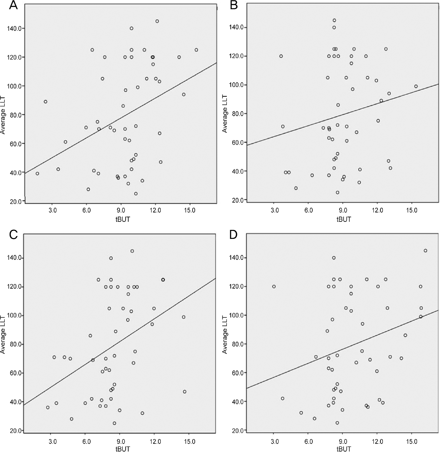 | Figure 1.Pearson correlation scatter plot of tear break-up time (tBUT) and average lipid layer thickness (LLT) using implanted data set 1 (A), 2 (B), 3 (C), and 4 (D). (A) r = 0.376, p-value = 0.008. (B) r = 0.220, p-value = 0.035. (C) r = 0.390, p-value = 0.006. (D) r = 0.276, p-value = 0.049. p-values were calculated by Pearson correlation test. |
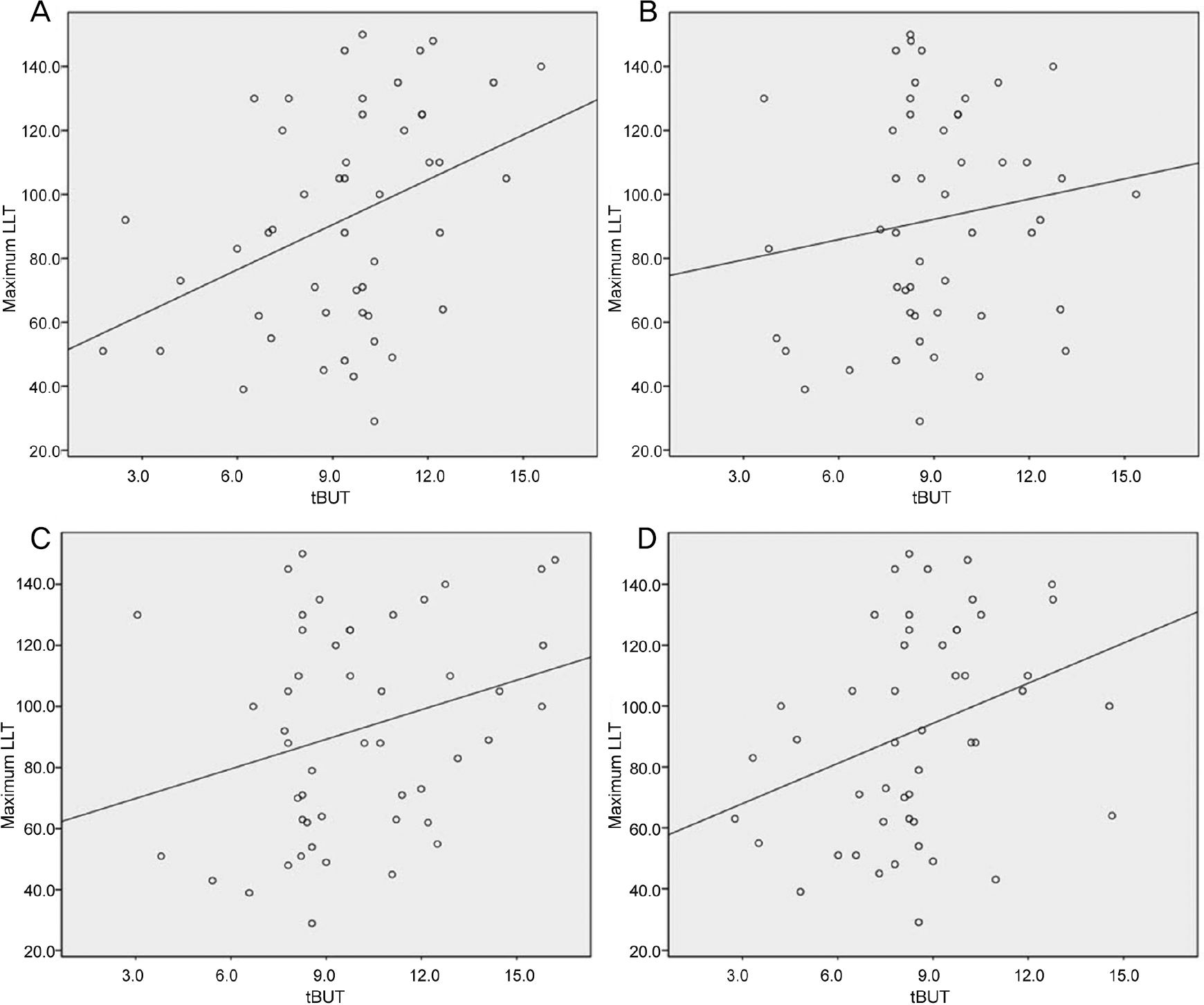 | Figure 2.Pearson correlation scatter plot of tear break-up time (tBUT) and maximum lipid layer thickness (LLT) using implanted data set 1 (A), 2 (B), 3 (C), and 4 (D). (A) r = 0.390, p-value = 0.006. (B) r = 0.178, p-value = 0.042. (C) r = 0.331, p-value = 0.020. (D) r = 0.279, p-value = 0.049. p-values were calculated by Pearson correlation test. |
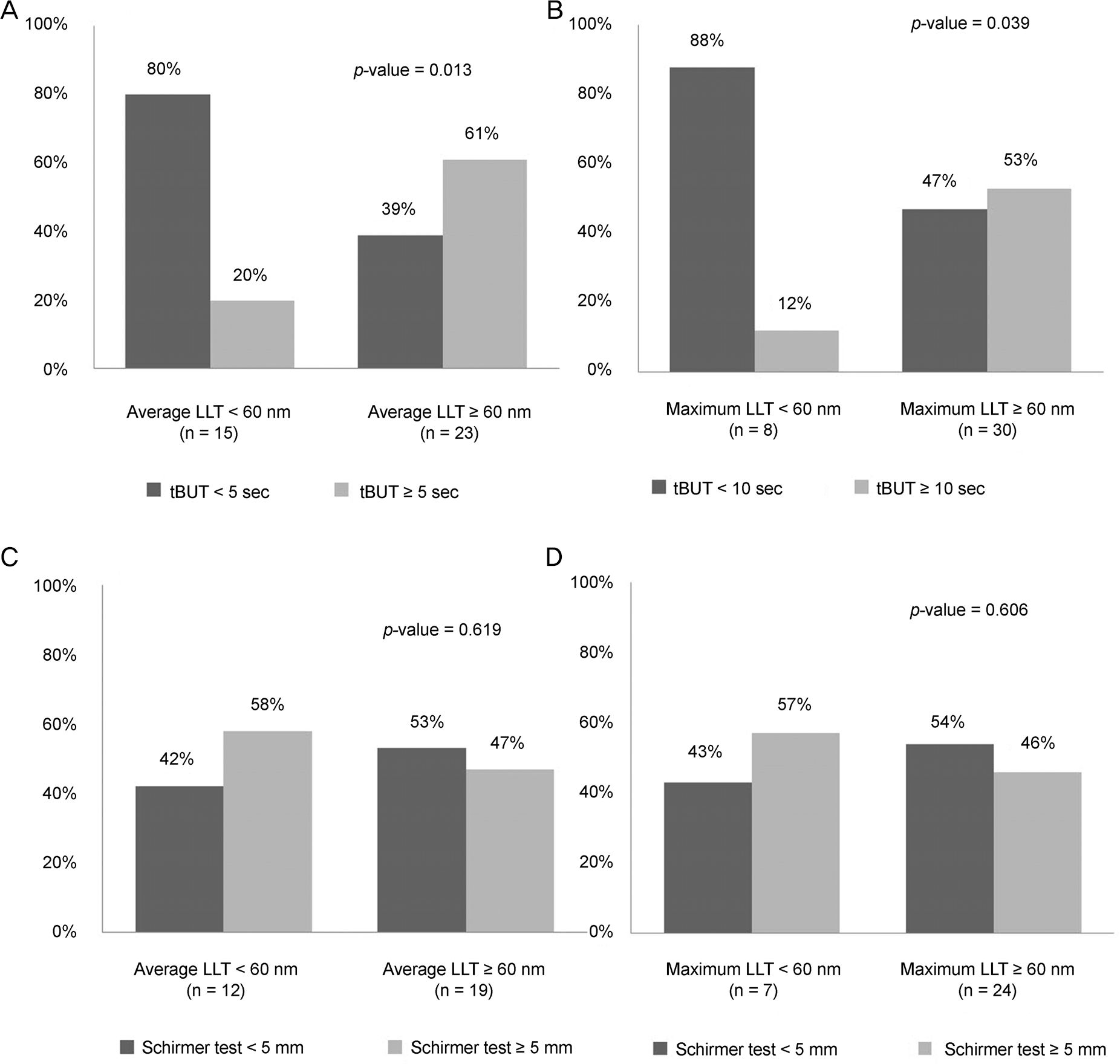 | Figure 3.The percentage of patients with shorten (<10 sec) or normal (≥10 sec) tear break-up time (tBUT) in each relatively thin lipid layer thickness (LLT) (<60 nm) or relatively thick LLT (≥60 nm) (A: average LLT, B: maximum LLT). The percentage of patients with shorten (<5 mm) or normal (≥5 mm) Schirmer test in each relatively thin LLT (<60 nm) or relatively thick LLT (≥60 nm) (C: average LLT, D: maximum LLT). p-values were calculated by Chi-square test. |
Table 1.
Clinical characteristics of patients
| Characteristics | Value (range*) |
|---|---|
| Age (years) | 48.15 ± 15.69 (18-72) |
| Sex (male:female) | 18:31 |
| Schirmer’s test (mm) | 8.45 ± 7.80 (2-20) |
| tBUT (sec) | 5.00 ± 3.43 (1-10) |
| Average LLT (nm) | 79.55 ± 37.31 (25-140) |
| Maximum LLT (nm) | 93.30 ± 37.11 (29-150) |
| Minimum LLT (nm) | 62.70 ± 32.33 (23-115) |
| Total blinking (times/20 sec) | 5.80 ± 3.24 (2-12) |
| Partial blinking (times/20 sec) | 3.30 ± 2.36 (0-9) |
| Partial blinking/total blinking ratio | 0.59 ± 0.31 (0-1) |
Table 2.
Correlation analysis among LLT and clinical factors using 4 sets of implantation data
| Data set | Average LLT | Maximum LLT | Minimum LLT | Difference‡ | Total blink | Partial blink | |
|---|---|---|---|---|---|---|---|
| Schirmer test | 1 | 0.149 | 0.050 | 0.229 | 0.361* | -0.383* | -0.157 |
| (0.307) | (0.733) | (0.114) | (0.011) | (0.007) | (-0.554) | ||
| 2 | 0.125 | 0.047 | 0.147 | 0.199 | -0.370† | -0.213† | |
| (0.221) | (0.646) | (0.147) | (0.005) | (0.000) | (0.009) | ||
| 3 | 0.044 | -0.031 | 0.056 | -0.191 | -0.356* | 0.170 | |
| (0.765) | (0.933) | (0.700) | (0.199) | (0.012) | (0.243) | ||
| 4 | 0.155 | 0.074 | 0.221 | -0.291* | -0.271 | -0.292* | |
| (0.297) | (0.615) | (0.128) | (0.043) | (0.060) | (0.042) | ||
| tBUT | 1 | 0.376† | 0.390† | 0.284* | -0.322* | -0.114 | -0.109 |
| (0.008) | (0.006) | (0.048) | (0.024) | (0.435) | (0.456) | ||
| 2 | 0.220* | 0.178* | 0.179 | 0.199 | 0.247* | -0.071 | |
| (0.035) | (0.042) | (0.090) | (0.051) | (0.014) | (0.499) | ||
| 3 | 0.390† | 0.331* | 0.254 | 0.249 | 0.245 | -0.044 | |
| (0.006) | (0.020) | (0.079) | (0.096) | (0.090) | (0.765) | ||
| 4 | 0.276* | 0.279* | 0.297* | 0.035 | -0.198 | -0.203 | |
| (0.049) | (0.049) | (0.039) | (0.913) | (0.177) | (0.162) | ||
| Staining grade | 1 | 0.148 | 0.186 | 0.035 | 0.364* | 0.160 | 0.123 |
| (0.311) | (0.202) | (0.811) | (0.101) | (0.582) | (0.399) | ||
| 2 | 0.221* | 0.195 | 0.252* | 0.206* | 0.196 | 0.213* | |
| (0.029) | (0.069) | (0.012) | (0.042) | (0.067) | (0.035) | ||
| 3 | 0.220 | 0.239 | 0.192 | 0.179 | 0.209 | 0.170 | |
| (0.129) | (0.099) | (0.211) | (0.217) | (0.152) | (0.243) | ||
| 4 | 0.220 | 0.290* | 0.152 | 0.383* | 0.169 | 0.264 | |
| (0.129) | (0.044) | (0.296) | (0.010) | (0.247) | (0.067) | ||
| LL MGD grade | -0.015 | -0.018 | 0.000 | -0.017 | -0.017 | 0.107 | |
| (0.916) | (0.959) | (0.999) | (0.906) | (0.909) | (0.465) |
Table 3.
Partial correlation analysis controlling for age and sex of the subject using 4 sets of implantation data
| Data set | Average LLT | Maximum LLT | Minimum LLT | Difference‡ | Total blink | Partial blink | |
|---|---|---|---|---|---|---|---|
| Schirmer test | 1 | 0.203 | 0.123 | 0.270 | -0.315* | -0.397* | -0.270 |
| (0.171) | (0.410) | (0.066) | (0.031) | (0.006) | (-0.067) | ||
| 2 | 0.133 | 0.049 | 0.150 | -0.222* | -0.370† | -0.300† | |
| (0.198) | (0.633) | (0.144) | (0.030) | (0.000) | (0.003) | ||
| 3 | 0.041 | -0.049 | 0.051 | -0.238 | -0.357* | 0.300* | |
| (0.784) | (0.743) | (0.733) | (0.108) | (0.014) | (0.041) | ||
| 4 | 0.164 | 0.102 | 0.241 | -0.301* | -0.287* | -0.218* | |
| (0.272) | (0.469) | (0.102) | (0.040) | (0.049) | (0.140) | ||
| tBUT | 1 | 0.396† | 0.396† | 0.286 | -0.320* | -0.114 | -0.208 |
| (0.006) | (0.006) | (0.051) | (0.028) | (0.445) | (0.162) | ||
| 2 | 0.309† | 0.273* | 0.188 | 0.241* | 0.246* | -0.069 | |
| (0.002) | (0.007) | (0.066) | (0.018) | (0.016) | (0.505) | ||
| 3 | 0.414† | 0.361* | 0.263 | 0.288 | 0.245 | -0.047 | |
| 4 | (0.004) 0.242* | (0.013)0.259* | (0.074)0.288 | (0.050)-0.020 | (0.097)-0.207 | (0.752)-0.136 | |
| (0.018) | (0.035) | (0.050) | (0.891) | (0.162) | (0.363) |
 XML Download