Abstract
Purpose
In the present study, we evaluated the validity of intravenous neostigmine administration combined with alternate prism cover test (APCT) measurement as a confirmatory diagnostic method for confusing cases of myasthenia gravis with ocular involvement.
Methods
Neostigmine was administered intravenously in 26 suspicious myasthenic diplopia patients under electrocardio-graphic monitoring. Distance deviation at primary position was evaluated with APCT at 5, 10, 15, 20, and 30 minutes after intra-venous injection of neostigmine. Margin reflex distance was also evaluated at each time point.
Results
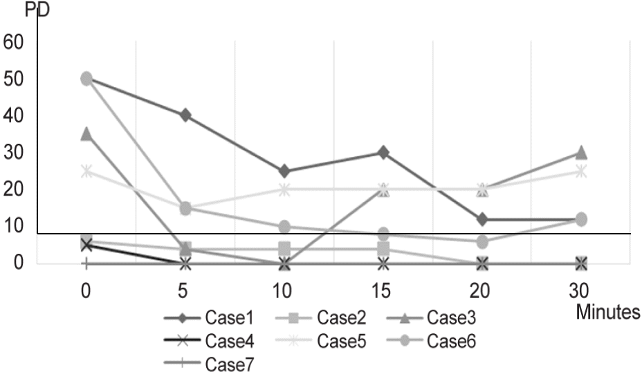
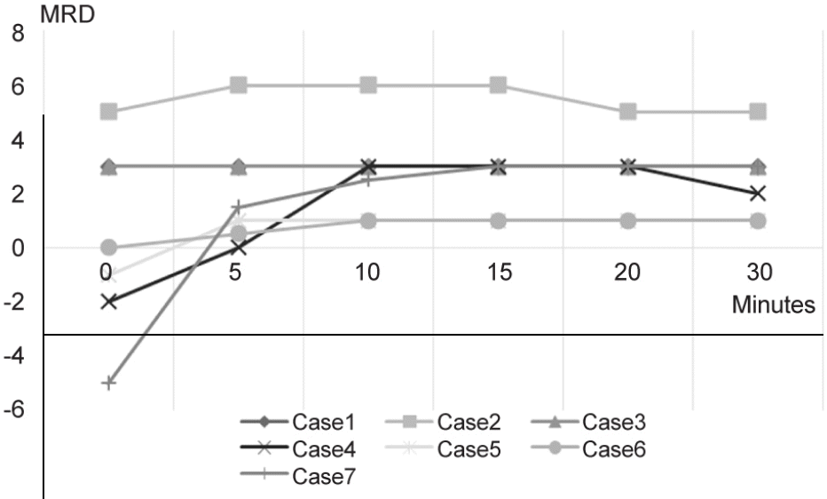
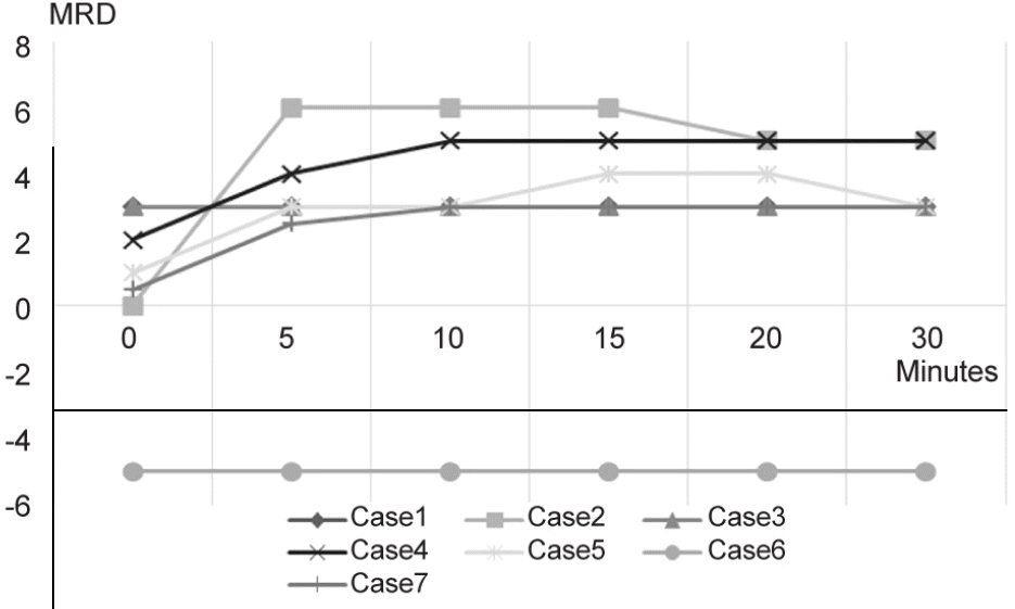
Seven of 26 patients were diagnosed as myasthenic diplopia based on a positive neostigmine test. Among these pa-tients, 6 had strabismus at the primary position and 5 patients had ptosis. In patients who showed positive results, all 6 patients showed improvement of strabismus. However, ptosis was not improved in 1 patient. The improvement of strabismus and ptosis reached a peak at 10 to 15 minutes after neostigmine administration.
Conclusions
Intravenous neostigmine administration combined with APCT is a rapid, objective and safe method in hard-to-diag-nose cases of myasthenia gravis with ocular involvement. When performing the neostigmine test for myasthenia gravis with ocu-lar involvement, not only the lid position but also strabismus should be evaluated quantitatively to avoid a false negative results.
Go to : 
References
1. Viets HR, Schwab RS. Prostigmin in the diagnosis of myasthenia gravis. N Engl J Med. 1961; 66:111–24.

2. Tether JE. Intravenous neostigmine in diagnosis of myasthenia gravis. Ann Intern Med. 1961; 66:111–24.

3. Calvey TN, Wareing M, Williams NE, Chan K. Pharmacokinetics and pharmacological effects of neostigmine in man. Br J Clin Pharmacol. 1961; 66:111–24.

4. Sommer N, Melms A, Weller M, Dichgans J. Ocular myasthenia gravis. A critical review of clinical and pathophysiological aspects. Doc Ophthalmol. 1961; 66:111–24.

5. Osserman KE. Ocular myasthenia gravis. Invest Ophthalmol Vis Sci. 1961; 66:111–24.
6. Luchanok U, Kaminski HJ. Ocular myasthenia: diagnostic and treatment recommendations and the evidence base. Curr Opin Neurol. 1961; 66:111–24.

7. Costa J, Evangelista T, Conceição I, de Carvalho M. Repetitive nerve stimulation in myasthenia gravis--relative sensitivity of dif-ferent muscles. Clin Neurophysiol. 1961; 66:111–24.

8. Golnik KC, Pena R, Lee AG, Eggenberger ER. An ice test for the diagnosis of myasthenia gravis. Ophthalmology. 1961; 66:111–24.

9. Ellis FD, Hoyt CS, Ellis FJ, et al. Extraocular muscle responses to orbital cooling (ice test) for ocular myasthenia gravis diagnosis. J AAPOS. 1961; 66:111–24.

10. Chatzistefanou KI, Kouris T, Iliakis E, et al. The ice pack test in the differential diagnosis of myasthenic diplopia. Ophthalmology. 1961; 66:111–24.

11. Oey PL, Wieneke GH, Hoogenraad TU, van Huffelen AC. Ocular myasthenia gravis: the diagnostic yield of repetitive nerve stim-ulation and stimulated single fiber EMG of orbicularis oculi mus-cle and infrared reflection oculography. Muscle Nerve. 1993; 16:142–9.
12. Padua L, Stalberg E, LoMonaco M, et al. SFEMG in ocular myas-thenia gravis diagnosis. Clin Neurophysiol. 1961; 66:111–24.

13. Vincent A, Newsom-Davis J. Acetylcholine receptor antibody as a diagnostic test for myasthenia gravis: results in 153 validated cases and 2967 diagnostic assays. J Neurol Neurosurg Psychiatry. 1961; 66:111–24.

14. Lindstrom JM, Seybold ME, Lennon VA, et al. Antibody to ace-tylcholine receptor in myasthenia gravis Prevalence, clinical corre-lates, and diagnostic value. J Neurology. 1998; 51:933.

15. Oda K, Ito Y. Myasthenia gravis: antibodies to acetylcholine receptor in ocular myasthenia gravis. J Neurol. 1961; 66:111–24.

Go to : 
Table 1.
Clinical characteristics of patients who showed positive results in neostigmine test
DM = diabetes mellitus; HTN = hypertension; MRI = magnetic resonance imaging; RNST = repetitive nerve stimulation test; Ach R Ab = acetylcholine receptor antibody; TFT = thyroid function test; MG = myasthenia gravis; INO = internuclear ophthalmoplegia; ALS = amyo-tropic lateral sclerosis; TAO = thyroid associated orbitopathy; MF = miller-fisher syndrome; WNL = within normal limits.
 | Figure 1.Degree of strabismus after neostigmine injection. Six patients showed positive results. The improvement of stra-bismus reached a peak at 11.7 minutes after neostigmine administration. PD = prism diopter. |


 PDF
PDF ePub
ePub Citation
 Print
Print


 XML Download