Abstract
Purpose
To determine the normal ranges of various indexes of the pupillary light reflex measured by automated pupillometry in Koreans.
Methods
We retrospectively analyzed 90 healthy adults who did not have any ocular diseases other than refractive errors. The direct pupillary light reflex was measured with an automated dynamic pupillometer (PLR-200, NeurOptics Inc., Irvine, CA, USA). A total of 7 indices were measured as follows; the maximum and minimum pupil diameters, constriction latency, constriction ratio, maximum constriction velocity, average constriction velocity and average dilation velocity.
Results
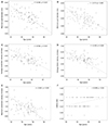
There were no significant differences in quantitative indexes of the pupillary light reflex between fellow eyes. A significant decrease in maximum pupil diameter, minimum pupil diameter, maximum constriction velocity, average constriction velocity and average dilation velocity were observed with aging. In contrast, a significant increase in constriction latency was observed with aging. There were no differences in quantitative pupil measurements according to gender (p<0.001).
Conclusions
Quantitative measurements of the pupillary light reflex by dynamic pupillometry showed no significant differences between fellow eyes. A significant decrease in pupil size, constriction velocity and dilation velocity, and an increase in pupil constriction latency were observed with aging.
Figures and Tables
 | Figure 1Correlations between age and quantitative measurements of the pupillary light reflex. The maximum pupil diameter (mm) (A), minimum pupil diameter (mm) (B), average constriction velocity (mm/sec) (C), average dilation velocity (mm/sec) (D) and maximum constriction velocity (mm/sec) (E) significantly decreased with age. In contrast, a significant increase in constriction latency (sec) (F) was observed with aging (r = Pearson' correlation coefficient). |
References
1. Boev AN, Fountas KN, Karampelas I, et al. Quantitative pupillometry: normative data in healthy pediatric volunteers. J Neurosurg. 2005; 103:6 Suppl. 496–500.
2. Miller NR, Subramanian PS, Patel VR. Walsh and Hyot's Clinical Neuro-Ophthalmology: The Essentials. 3rd ed. Baltimore: Wolters Kluwer;2016. p. 264–268.
3. Lee IB, Choi BH, Mun YS, Hwang JM. Relative afferent pupillary defect in normal subjects in 10 to 39 years of age. J Korean Ophthalmol Soc. 2005; 46:1034–1036.
4. Zafar SF, Suarez JI. Automated pupillometer for monitoring the critically ill patient: a critical appraisal. J Crit Care. 2014; 29:599–603.
5. Meeker M, Du R, Bacchetti P, et al. Pupil examination: validity and clinical utility of an automated pupillometer. J Neurosci Nurs. 2005; 37:34–40.
6. Martínez-Ricarte F, Castro A, Poca MA, et al. Infrared pupillometry. Basic principles and their application in the non-invasive monitoring of neurocritical patients. Neurologia. 2013; 28:41–51.
7. Morris GF, Juul N, Marshall SB, et al. Executive Committee of the International Selfotel Trial. Neurological deterioration as a potential alternative endpoint in human clinical trials of experimental pharmacological agents for treatment of severe traumatic brain injuries. Neurosurgery. 1998; 43:1369–1372. discussion 1372-4.
8. Litvan I, Saposnik G, Mauriño J, et al. Pupillary diameter assessment: need for a graded scale. Neurology. 2000; 54:530–531.
9. Ko BU, Ryu WY, Park WC. Pupil size in the normal Korean population according to age and illuminance. J Korean Ophthalmol Soc. 2011; 52:401–406.
10. Martinez CE, Applegate RA, Klyce SD, et al. Effect of pupillary dilation on corneal optical aberrations after photorefractive keratectomy. Arch Ophthalmol. 1998; 116:1053–1062.
11. Murray RB, Loughnane MH. Infrared video pupillometry: a method used to measure the pupillary effects of drugs in small laboratory animals in real time. J Neurosci Methods. 1981; 3:365–375.
12. Roberts CW, Koester CJ. Optical zone diameters for photorefractive corneal surgery. Invest Ophthalmol Vis Sci. 1993; 34:2275–2281.
13. Taylor WR, Chen JW, Meltzer H, et al. Quantitative pupillometry, a new technology: normative data and preliminary observations in patients with acute head injury. Technical note. J Neurosurg. 2003; 98:205–213.
14. Schallenberg M, Bangre V, Steuhl KP, et al. Comparison of the Colvard, Procyon, and Neuroptics pupillometers for measuring pupil diameter under low ambient illumination. J Refract Surg. 2010; 26:134–143.
15. Baek JS, Park JH, Yoo ES, et al. Comparison of Colvardpupillometer, ORBScan II and Sirius in determining pupil size for refractive surgery. J Korean Ophthalmol Soc. 2013; 54:1175–1179.
16. Du R, Meeker M, Bacchetti P, et al. Evaluation of the portable infrared pupillometer. Neurosurgery. 2005; 57:198–203.
17. Lee TJ, Kim HS, Jung JW, et al. Comparison of automatic pupillometer and pupil card for measuring pupil size. J Korean Ophthalmol Soc. 2015; 56:863–867.
18. Couret D, Boumaza D, Grisotto C, et al. Reliability of standard pupillometry practice in neurocritical care: an observational, double-blinded study. Crit Care. 2016; 20:99.
19. Chesnut RM, Gautille T, Blunt BA, et al. The localizing value of asymmetry in pupillary size in severe head injury: relation to lesion type and location. Neurosurgery. 1994; 34:840–845. discussion 845-6.
20. Manley GT, Larson MD. Infrared pupillometry during uncal herniation. J Neurosurg Anesthesiol. 2002; 14:223–228.
21. Hsieh YT, Hu FR. The correlation of pupil size measured by Colvard pupillometer and Orbscan II. J Refract Surg. 2007; 23:789–795.
22. Nakamura K, Bissen-Miyajima H, Oki S, Onuma K. Pupil sizes in different Japanese age groups and the implications for intraocular lens choice. J Cataract Refract Surg. 2009; 35:134–138.
23. Libby WL Jr, Lacey BC, Lacey JI. Pupillary and cardiac activity during visual attention. Psychophysiology. 1973; 10:270–294.


 PDF
PDF ePub
ePub Citation
 Print
Print


 XML Download