Journal List > J Korean Ophthalmol Soc > v.58(6) > 1010780

Kim and Son: Evaluation of Bony Nasolacrimal Ducts in Koreans with Primary Acquired Nasolacrimal Duct Obstruction

Abstract

Purpose

To evaluate the morphometric differences of bony nasolacrimal ducts (BNLDs) using computed tomography (CT) in Koreans with primary acquired nasolacrimal duct obstruction (PANDO).

Methods

From March 2014 to March 2016, 40 unilateral PANDO patients and 40 control subjects were retrospectively reviewed. The axial, sagittal, and coronal planes of CT were used for image evaluation. The proximal, minimal, and distal transverse diameters (TDs) of the BNLD were assessed. The length, sagittal orientation angle of BNLD, relative lacrimal sac-BNLD angle, nasal floor-BNLD angle, and turbinate angle were investigated. In addition, the distance between the bilateral BNLD and inter-frontozygomatic suture distance were also measured.

Results

There were no morphologic differences between the PANDO and non-PANDO sides within PANDO patients. The proximal and minimum BNLD TD measurements were significantly narrower in the PANDO patients, as compared with the control group (p = 0.010 and p = 0.017, respectively). The lacrimal sac-BNLD angle, nasal floor-BNLD angle, and turbinate angle also exhibited statistically significant differences between the PANDO patients and the control group (p = 0.019, p = 0.001, and p < 0.001, respectively).

Conclusions

Although this study was performed in a small group, the narrow proximal and minimum BNLD TD in PANDO patients (in both the PANDO and non-PANDO sides) may be associated with PANDO development in Koreans. Additionally, the narrow lacrimal sac-BNLD, nasal floor-BNLD, and turbinate angle might be possible causative factors of PANDO.

Figures and Tables

Figure 1

Bony nasolacrimal duct (BNLD) measurements with computed tomography (CT). (A) Axial CT image in bone window demonstrates BNLD transverse diameter (yellow line) measurement. (B) It reveals distance between the proximal BNLD (green line). (C) It shows inter-frontozygomatic suture distance (white line).
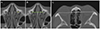
jkos-58-634-g001
Figure 2

Bony nasolacrimal duct (BNLD) measurements with computed tomography (CT). (A) Sagittal CT image reveals BNLD length (orange line) and proximal, minimal, distal BNLD anteroposterior diameter (yellow lines) measurements, respectively. ‘a’ demonstrates measurement of sagittal orientation angle between long axis (white line) of BNLD and nasal floor (black line). (B) Coronal CT image shows relative lacrimal sac-BNLD orientation angle ‘b’ between long axis of lacrimal sac fossa (green line) and BNLD (white line). ‘c’ reveals angle between long axis of BNLD (white line) and nasal floor (black line). (C) It demonstrates angle (red line) between the bony inferior turbinate and upper part of the medial wall of the maxillary sinus.
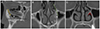
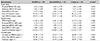
jkos-58-634-g002
Table 1

Patient characteristics

jkos-58-634-i001

Values are presented as mean ± SD unless otherwise indicated.

PANDO = primary acquired nasolacrimal duct obstruction; SD = standard deviation; M = male; F = female; OD = oculus dexter; OS = oculus sinister.

*No statistical significance using a student t-test; No statistical significance using a χ2 for independence (p < 0.05).
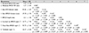
Table 2

Values of measurements in PANDO side, non-PANDO side, and control groups

jkos-58-634-i002

Values are presented as mean ± SD unless otherwise indicated.

PANDO = primary acquired nasolacrimal duct obstruction; BNLD = bony nasolacrimal duct; TD = transverse diameter; FZS = frontozygomatic suture; APD = anteroposterior diameter; SD = standard deviation.

*One way analysis of variance (ANOVA) test (p < 0.05). There was no statistically significant difference between PANDO side and non-PANDO side (p > 0.5, Post hoc test).

Table 3

Pearson correlation coefficients between values of measurements

jkos-58-634-i003

Values are presented as mean ± SD unless otherwise indicated.

BNLD = bony nasolacrimal duct; TD = transverse diameter; FZS = frontozygomatic suture; SD = standard deviation.

*Pearson correlation coefficient (r): p < 0.05; Pearson correlation coefficient (r): p < 0.01.

References

1. Linberg JV, McCormick SA. Primary acquired nasolacrimal duct obstruction. A clinicopathologic report and biopsy technique. Ophthalmology. 1986; 93:1055–1063.
2. Ohtomo K, Ueta T, Toyama T, Nagahara M. Predisposing factors for primary acquired nasolacrimal duct obstruction. Graefes Arch Clin Exp Ophthalmol. 2013; 251:1835–1839.
3. Seider N, Miller B, Beiran I. Topical glaucoma therapy as a risk factor for nasolacrimal duct obstruction. Am J Ophthalmol. 2008; 145:120–123.
4. Bulbul E, Yazici A, Yanik B, et al. Morphometric evaluation of bony nasolacrimal canal in a Caucasian population with primary acquired nasolacrimal duct obstruction: A Multidetector Computed Tomography Study. Korean J Radiol. 2016; 17:271–276.
5. Gul A, Aslan K, Karli R, et al. A possible cause of nasolacrimal duct obstruction: narrow angle between inferior turbinate and upper part of the medial wall of the maxillary sinus. Curr Eye Res. 2016; 41:729–733.
6. Janssen AG, Mansour K, Bos JJ, Castelijns JA. Diameter of the bony lacrimal canal: normal values and values related to nasolacrimal duct obstruction: assessment with CT. AJNR Am J Neuroradiol. 2001; 22:845–850.
7. McCormick A, Sloan B. The diameter of the nasolacrimal canal measured by computed tomography: gender and racial differences. Clin Exp Ophthalmol. 2009; 37:357–361.
8. Shigeta K, Takegoshi H, Kikuchi S. Sex and age differences in the bony nasolacrimal canal: an anatomical study. Arch Ophthalmol. 2007; 125:1677–1681.
9. Estes JL, Tsiouris AJ, Christos PJ, Lelli GJ. Three-dimensional volumetric assessment of the nasolacrimal duct in patients with obstruction. Ophthal Plast Reconstr Surg. 2015; 31:211–214.
10. George M, Phillips CI. Epiphora and the bony naso-lacrimal canal. Br J Ophthalmol. 1956; 40:673–680.
11. Fasina O, Ogbole GI. CT assessment of the nasolacrimal canal in a black African Population. Ophthal Plast Reconstr Surg. 2013; 29:231–233.
12. Groessl SA, Sires BS, Lemke BN. An anatomical basis for primary acquired nasolacrimal duct obstruction. Arch Ophthalmol. 1997; 115:71–74.
13. Post RH. Tear duct size differences of age, sex and race. Am J Phys Anthropol. 1969; 30:85–88.
14. Takahashi Y, Kakizaki H, Nakano T. Bony nasolacrimal duct entrance diameter: gender difference in cadaveric study. Ophthal Plast Reconstr Surg. 2011; 27:204–205.
15. Takahashi Y, Nakamura Y, Nakano T, et al. The narrowest part of the bony nasolacrimal canal: an anatomical study. Ophthal Plast Reconstr Surg. 2013; 29:318–322.
16. Ramey NA, Hoang JK, Richard MJ. Multidetector CT of nasolacrimal canal morphology: normal variation by age, gender, and race. Ophthal Plast Reconstr Surg. 2013; 29:475–480.
17. Lee H, Ha S, Lee Y, et al. Anatomical and morphometric study of the bony nasolacrimal canal using computed tomography. Ophthalmologica. 2012; 227:153–159.
18. Takahashi Y, Nakata K, Miyazaki H, et al. Comparison of bony nasolacrimal canal narrowing with or without primary acquired nasolacrimal duct obstruction in a Japanese population. Ophthal Plast Reconstr Surg. 2014; 30:434–438.
19. Yong AM, Zhao DB, Siew SC, et al. Assessment of bony nasolacrimal parameters among Asians. Ophthal Plast Reconstr Surg. 2014; 30:322–327.
TOOLS
Similar articles