

 PDF
PDF ePub
ePub Citation
 Print
Print


Abstract
Case summary
A 48-year-old woman visited our ophthalmology department with decreased vision and disturbance of the visual field in the right eye, which began 2 days prior to presentation. The patient history indicated she had undergone total thyroidectomy 3 months prior and was given an oral calcium preparation. She had no eye pain, headache, tinnitus or diplopia. Her best corrected visual acuity of both eyes was 1.0, and color vision was normal in both eyes although a mild relative afferent pupillary defect was present in the right eye. Severe bilateral optic disc edema was present in the right eye. A Humphrey visual field test revealed an enlarged blind spot and peripheral nasal step scotoma in the right eye. The Cerebrospinal fluid (CSF) opening pressure was within the normal range and there were no abnormal findings regarding CSF. Additionally, there were no remarkable findings on brain magnetic resonance imaging nor neurologic tests. Her serum calcium was 5.9 mg/dL (normal range: total calcium 8.7-10.6 mg/dL), and an intravenous calcium supplement was started. Visual disturbance and optic disc edema improved 2 days after replacement and the optic disc edema completely dissolved 2 months later.
Figures and Tables
Figure 1
Humphrey visual field on the initial examination. The Blind spot was enlarged and a scotoma was noticeable in the nasal visual field of the right eye.
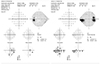
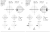
Figure 2
Fundus photographs on the initial examination. There were optic disc swellings in both eyes. Optic disc swelling was asymmetric and more severe in the right eye. The right retinal vein was tortuous.


References
1. Van Stavern GP. Optic disc edema. Semin Neurol. 2007; 27:233–243.
2. Lyle DJ. The ocular syndrome of cataract and papilledema in the manifest form of parathyroid deficiency. Am J Ophthalmol. 1948; 31:580–584.
3. Sheldon RS, Becker WJ, Hanley DA, Culver RL. Hypoparathyroidism and pseudotumor cerebri: an infrequent clinical association. Can J Neurol Sci. 1987; 14:622–625.
4. Mor F, Wysenbeek AJ. Evidence on computed tomography of pseudotumour cerebri in hypoparathyroidism. Br J Radiol. 1988; 61:158–160.
5. Palmer RF, Searles HH, Boldrey EB. Papilloedema and hypoparathyroidism simulating brain tumor. J Neurosurg. 1959; 16:378–384.
6. Friedman DI. Papilledema and pseudotumor cerebri. Ophthalmol Clin North Am. 2001; 14:129–147.
7. Goyal JL, Kang J, Gupta R, et al. Bilateral papilledema in hypocalcemia. Delhi J ophthalmol. 2012; 23:127–130.
8. McLean C, Lobo R, Brazier DJ. Optic disc involvement in hypocalcaemia with hypoparathyroidism: papilloedema or optic neuropathy? Neuroophthalmology. 1998; 20:117–124.
9. Abu-Ain M, Aazem S, Morton C, et al. A rare potentially treatable cause of bilateral optic disc swelling. BMJ Case Rep. 2010; 2010:pii: bcr0320102835.
10. Bajandas FJ, Smith JL. Optic nueritis in hypoparathyroidism. Neurology. 1976; 26:451–454.
11. Nathanson JA. Beta-Adrenergic-sensitive adenylate cyclase in choroid plexus: properties and cellular localization. Mol Pharmacol. 1980; 18:199–209.
12. Sambrook MA, Hill LF. Cerebrospinal fluid absorption in primary hypoparathyroidism. J Neurol Neurosurg Psychiatry. 1977; 40:1015–1017.
13. Breuer AC, Atkinson MB. Calcium dependent modulation of fast axonal transport. Cell Calcium. 1988; 9:293–301.
 XML Download