

 PDF
PDF ePub
ePub Citation
 Print
Print


Abstract
Case summary
A 10-year-old boy visited our pediatric genetic metabolic clinic for evaluation of his overall developmental delay and short stature. The boy was diagnosed with mucolipidosis type II (I-cell disease) using plasma enzyme assay and DNA sequencing of the GNPTAB gene mutation. An ophthalmologic investigation was then performed, and a depressed nasal bridge, broad nose, and swelling in the upper lid of both eyes were noted. The best corrected visual acuity was 0.32 and 0.1 and the intraocular pressure was 35 mmHg and 24 mmHg in the right and left eyes, respectively. The anterior chamber angles of both eyes were normal and mild cornea opacity in both eyes was observed. Fundus examination revealed retinal atrophy with folds in both eyes, as well as optic disc edema and optic atrophy in the right and left eyes, respectively. Atherosclerotic changes in the retinal vessels and cystoid macular edema in the left eye were observed, and ocular ultrasound revealed increased posterior sclera thickness in both eyes.
Conclusions
Ocular manifestations of mucolipidosis type II are not currently well-known, and differentiation from other metabolic disorders may be difficult. An ophthalmic work-up can assist in diagnosis, and regular ophthalmic examinations should be used to maintain visual function in mucolipidosis patients.
Figures and Tables
Figure 1
Photograph of the patient's face. Depressed nasal bridge and upper eyelid swelling in both eyes were observed.

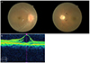
Figure 2
The fundus photographs and the optical coherence tomography of the patient. (A) Fundus examination of the both eyes showed retinal atrophy with retinal fold (white arrow). Optic disc edema in right eye and optic disc atrophy in left eye were observed. Left eye showed retinal artery narrowing with atherosclerotic changes (black arrows) and cystoid macular edema. (B) The optical coherence tomography showed cystoid macular edema in left eye.
References
1. Libert J, Van Hoof F, Farriaux JP, Toussaint D. Ocular findings in I-cell disease (mucolipidosis type II). Am J Ophthalmol. 1977; 83:617–628.
2. Kudo M, Brem MS, Canfield WM. Mucolipidosis II (I-cell disease) and mucolipidosis IIIA (classical pseudo-hurler polydystrophy) are caused by mutations in the GlcNAc-phosphotransferase alpha / beta -subunits precursor gene. Am J Hum Genet. 2006; 78:451–463.
3. Yano S, Moseley K, Wong L, et al. Glycosaminoglycan metabolism defects and atherosclerosis: frequent association of endothelial dysfunction in patients with Mucopolysaccharidosis. J Inherit Metab Dis. 2014; 37:255–261.
4. Ganesh A, Bruwer Z, Al-Thihli K. An update on ocular involvement in mucopolysaccharidoses. Curr Opin Ophthalmol. 2013; 24:379–388.
5. Fahnehjelm KT, Ashworth JL, Pitz S, et al. Clinical guidelines for diagnosing and managing ocular manifestations in children with mucopolysaccharidosis. Acta Ophthalmol. 2012; 90:595–602.
6. Ferrari S, Ponzin D, Ashworth JL, et al. Diagnosis and management of ophthalmological features in patients with mucopolysaccharidosis. Br J Ophthalmol. 2011; 95:613–619.
7. Olsen TW, Aaberg SY, Geroski DH, Edelhauser HF. Human sclera: thickness and surface area. Am J Ophthalmol. 1998; 125:237–241.
 XML Download