Journal List > J Korean Ophthalmol Soc > v.58(5) > 1010769

Kim and Kang: Endoscopic and Transconjunctival versus Transcaruncular and Transconjunctival Reconstruction of Medial and Inferior Orbital Wall Fractures

Abstract

Purpose

To compare two combined surgical techniques, endoscopic endonasal surgery with transconjunctival reconstruction and transcaruncular surgery with transconjunctival reconstruction, when used to treat both medial and inferior orbital wall fractures.

Methods

A retrospective review of 63 patients who were followed up from January 2011 to December 2014 at Inha University Hospital for surgical reconstruction of combined medial and inferior orbital wall fractures was undertaken. We compared between the patients the computed tomographic scans, diplopia, extraocular muscle (EOM) movements, and Hertel's exophthalmometer exams pre- and post-6 months surgery. A total of 29 patients received endoscopic transnasal surgery with trasconjunctival reconstruction, and 34 received transcaruncular surgery with trans-conjunctival reconstruction.

Results

There were no significant differences between the two combined methods in terms of the primary and peripheral gaze diplopia or the restriction of EOM movement 6 months after surgery. However, statistically significant differences were observed in exophthalmometer measurements 6 months after surgery.

Conclusions

The two combined surgical methods showed similar results in terms of postoperative primary and peripheral gaze diplopia, EOM restriction, and enophthalmos. With respect to postoperative peripheral diplopia, endoscopic endosnasal surgery with transconjunctival reconstruction showed several advantages over the other method considered in this study. An appropriate surgical method should be selected by comparing the relative advantages and disadvantages.

Figures and Tables

Figure 1

Clinical photographs of preoperative combined medial and inferior orbital wall fracture. (A) In near face view. (B) Worm's eye view, photographs of postoperative combined medial and inferior orbital wall fracture with Endoscopic transnasal and transconjunctival approach. (C) In near face view. (D) Worm's eye view.

jkos-58-579-g001
Figure 2

Orbital Computed tomography of the patient. Orbital Computed tomography of preoperative combined medial and inferior orbital wall fracture. (A) Axial view. (B) Coronal view. Well-reconstructed medial and inferior orbital wall fracture with Endoscopic transnasal and transconjunctival approach. (C) Axial view. (D) Coronal view.
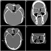
jkos-58-579-g002
Table 1

Demographics of the surgical patients

jkos-58-579-i001

Values are presented as mean ± SD unless otherwise indicated. Independent samples t-test was used to examine statistical difference.

Endoscopic with transconjunctival = endoscopic endonasal with transconjunctival reduction; Transcaruncular with transconjunctival = transcaruncular with transconjunctival reduction.

Table 2

Incidence of diplopia before and 6 months after repair

jkos-58-579-i002

Values are presented as n (%) unless otherwise indicated. Chi-Square test and Fisher's Exact test was used to examine statistical difference.

Endoscopic with transconjunctival = endoscopic endonasal with transconjunctival reduction; Transcaruncular with transconjunctival = transcaruncular with transconjunctival reduction; Pre op = preoperative; Postop = postoperative.

Table 3

Extraocular muscle restriction before and 6 months after surgical repair

jkos-58-579-i003

Values are presented as n (%) unless otherwise indicated. Chi-Square test and Fisher's Exact test was used to examine statistical difference.

Endoscopic = endoscopic endonasal reduction; Transcaruncular = transcaruncular reduction; Preop = preoperative; Postop = postoperative.

Table 4

Hertel's exophthalmometer measure before and 6 months after surgical repair

jkos-58-579-i004

Values are presented as mean ± SD unless otherwise indicated. Independent samples t-test was used to examine statistical difference.

Endoscopic = endoscopic endonasal reduction; Transcaruncular = transcaruncular reduction; Preop Exo = preoperative exophthalmometer measure; Postop 6 month Exo = postoperative 6 months exophthalmometer measure; Preop Exo diff = preoperative exophthalmometer measurement difference; Postop 6 month Exo diff = postoperative 6 months exophthalmometer measurement difference.

References

1. Ahn SK, Jung SW. The clinical aspects of orbital fractures proven by computed tomography. J Korean Ophthalmol Soc. 1997; 38:2077–2083.
2. Lee SY, Kim SY, Kim HB. Orbital fractures evaluated by computed tomography. J Korean Ophthalmol Soc. 1990; 31:249–253.
3. Dodick JM, Galin MA, Littleton JT, Sod LM. Concomitant medial wall fracture and blowout fracture of the orbit. Arch Ophthalmol. 1971; 85:273–276.
4. Nolasco FP, Mathog RH. Medial orbital wall fractures: classification and clinical profile. Otolaryngol Head Neck Surg. 1995; 112:549–556.
5. Warwar RE, Bullock JD, Ballal DR, Ballal RD. Mechanisms of orbital floor fractures: a clinical, experimental, and theoretical study. Ophthal Plast Reconstr Surg. 2000; 16:188–200.
6. Rhee JS, Kilde J, Yoganadan N, Pintar F. Orbital blowout fractures: experimental evidence for the pure hydraulic theory. Arch Facial Plast Surg. 2002; 4:98–101.
7. Jank S, Schuchter B, Emshoff R, et al. Clinical signs of orbital wall fractures as a function of anatomic location. Oral Surg Oral Med Oral Pathol Oral Radiol Endod. 2003; 96:149–153.
8. Burm JS, Chung CH, Oh SJ. Pure orbital wall fracture: new concepts and importance of medial orbital blowout fracture. Plast Reconstr Surg. 1999; 103:1839–1849.
9. Suhk JH, Ji SY, Kim TB, Yang WS. Internal fixation of Medpor(R) implant for prevention of enophthalmos in posteriorly extended orbital floor fracture. J Korean Cleft Palate-Craniofac Assoc. 2008; 9:55–61.
10. Wilkins RB, Havins WE. Current treatment of blow-out fractures. Ophthalmology. 1982; 89:464–466.
11. Lee MS, Lew H, Lee SY. The results of delayed repair for orbital wall fracture. J Korean Ophthalmol Soc. 1998; 39:1049–1054.
12. Kroll M, Wolper J. Orbital blowout fractures. Am J Ophthalmol. 1967; 64:1169–1172.
13. De Man K, Wijingaarde R, Hes J, de Jong PT. Influence of age on the management of blow-out fractures of the orbital floor. Int J Oral Maxillofac Surg. 1991; 20:330–336.
14. Hawes MJ, Dortzbach RK. Surgery on orbital floor fractures. Influence of time of repair and fracture size. Ophthalmology. 1983; 90:1066–1070.
15. Dulley B, Fells P. Long-term follow-up of orbital blow-out fracture with and without surgery. Mod Probl Ophthalmol. 1975; 14:467–470.
16. Emery JM, Noorden GK, Sclernitzauer DA. Orbital floor fractures: long-term follow-up of cases with and without surgical repair. Trans Am Acad Ophthalmol Otolaryngol. 1971; 75:802–812.
17. Mohadjer Y, Hartstein ME. Endoscopic orbital fracture repair. Otolaryngol Clin North Am. 2006; 39:1049–1057. viii
18. Rhee JS, Chen CT. Endoscopic approach to medial orbital wall fractures. Facial Plast Surg Clin North Am. 2006; 14:17–23.
19. Han K, Choi JH, Choi TH, et al. Comparison of endoscopic endonasal reduction and transcaruncular reduction for the treatment of medial orbital wall fractures. Ann Plast Surg. 2009; 62:258–264.
20. Lee CS, Yoon JS, Lee SY. Combined transconjunctival and transcaruncular approach for repair of large medial orbital wall fractures. Arch Ophthalmol. 2009; 127:291–296.
TOOLS
Similar articles