

 PDF
PDF ePub
ePub Citation
 Print
Print


Abstract
Purpose
To investigate the clinical features and surgical outcomes of rhegmatogenous retinal detachment (RRD) requiring sur-gery according to age.
Methods
Medical records of patients who underwent surgery for primary RRD between January 2008 and March 2016 were re-viewed retrospectively. Patients were classified into two groups according to age at diagnosis: the under-40 group and the over-40 group. The two groups were compared in terms of demographic features, ocular manifestation, operating methods, pri-mary anatomical success rate, and visual outcome.
Results
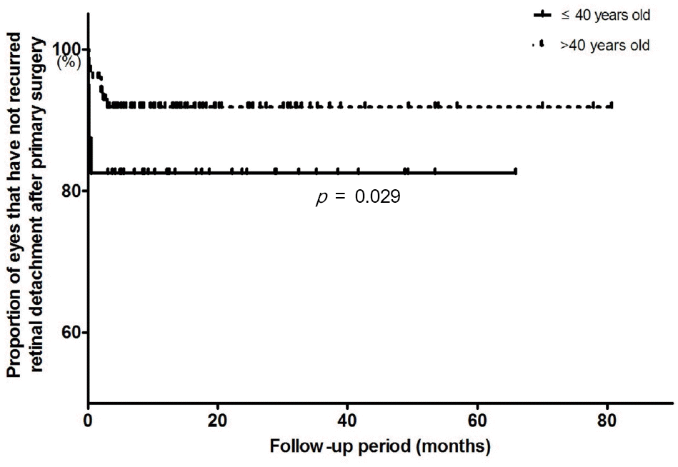
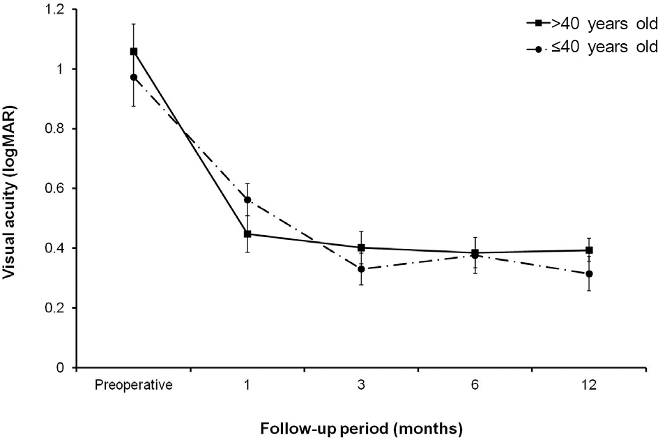
One hundred and forty-four eyes from 144 patients were included. Mean subject age was 48.6 ± 16.9 years old. The un-der-40 group involved 42 eyes from 42 patients, and the over-40 group included 102 eyes from 102 patients. Symptom duration was shorter in the under-40 group compared to the over-40 group (7.6 ± 10.7 days vs. 14.5 ± 24.4 days; p = 0.029). Proliferative vitreoretinopathy (PVR) occurred more frequently in the under-40 group (40.0% vs. 17.4%, p = 0.007) than in the over-40 group. The anatomical success rate of primary surgery was significantly different between the two groups; 78.6% in the under-40 group and 91.2% in the over-40 group ( p = 0.038). Preoperative PVR increased the rate of anatomical failure (40.0% vs. 6.2%, p < 0.001). The visual outcomes were not significantly different between the two groups.
References
1. Kuhn F, Aylward B. Rhegmatogenous retinal detachment: a re-appraisal of its pathophysiology and treatment. Ophthalmic Res. 1961; 66:111–24.

2. Heimann H, Bartz-Schmidt KU, Bornfeld N, et al. Scleral buckling versus primary vitrectomy in rhegmatogenous retinal detachment: a prospective randomized multicenter clinical study. Ophthalmology. 1961; 66:111–24.
3. Li X. Beijing Rhegmatogenous Retinal Detachment Study Group. Incidence and epidemiological characteristics of rhegmatogenous retinal detachment in Beijing, China. Ophthalmology. 2003; 110:2413–7.
4. Wilkes SR, Beard CM, Kurland LT, et al. The incidence of retinal detachment in Rochester, Minnesota, 1970-1978. Am J Ophthalmol. 1961; 66:111–24.
5. Törnquist R, Stenkula S, Törnquist P. Retinal detachment. A study of a population-based patient material in Sweden 1971-1981. I. Epidemiology. Acta Ophthalmol (Copenh). 1961; 66:111–24.
6. Chen SN, Lian IB, Wei YJ. Epidemiology and clinical character-istics of rhegmatogenous retinal detachment in Taiwan. Br J Ophthalmol. 1961; 66:111–24.
7. Choi SW, Kim KS, Kim YC. Clinical characteristics of rhegmatog-enous retinal detachment in patients under 40 years of age. J Korean Ophthalmol Soc. 1961; 66:111–24.
8. Sasaki K, Ideta H, Yonemoto J, et al. Epidemiologic characteristics of rhegmatogenous retinal detachment in Kumamoto, Japan. Graefes Arch Clin Exp Ophthalmol. 1961; 66:111–24.
9. Polkinghorne PJ, Craig JP. Northern New Zealand Rhegmatogenous Retinal Detachment Study: epidemiology and risk factors. Clin Experiment Ophthalmol. 1961; 66:111–24.
10. Park SJ, Choi NK, Park KH, Woo SJ. Five year nationwide in-cidence of rhegmatogenous retinal detachment requiring surgery in Korea. PLoS One. 2013; 8:e80174.
11. Lee JW, Song SJ, Park YH. Clinical features and surgical results of rhegmatogenous retinal detachment in children. J Korean Ophthalmol Soc. 1961; 66:111–24.
12. Machemer R, Aaberg TM, Freeman HM, et al. An updated classi-fication of retinal detachment with proliferative vitreoretinopathy. Am J Ophthalmol. 1961; 66:111–24.
13. Byon IS, Pak KY, Lee SM, et al. Lens-save versus phacoemulsifi-cation with intraocular lens implantation in primary vitrectomy for phakic rhegmatogenous retinal detachment. J Korean Ophthalmol Soc. 1961; 66:111–24.
14. Chung H, Lee JH. Clnical analysis of retinal detachment. J Korean Ophthalmol Soc. 1961; 66:111–24.
15. Jeong SK, Park YG, Lee MK. A clinical study on rhegmatogenous retinal detachment. J Korean Ophthalmol Soc. 1961; 66:111–24.
16. Schepens CL, Marden D. Data on the natural history of retinal detachment. Further characterization of certain unilateral non-traumatic cases. Am J Ophthalmol. 1961; 66:111–24.
17. Chignell AH. Retinal detachment surgery without drainage of sub-retinal fluid. Am J Ophthalmol. 1961; 66:111–24.
18. Park JL, Kim SD, Yun IH. A clinical study of the rhegmatogenous retinal detachment. J Korean Ophthalmol Soc. 1961; 66:111–24.
19. Kang SM, Yoon SW, Chin HS, Moon YS. Factors affecting the vis-ual outcome after scleral buckle in rhegmatogenous retinal detach-ment involving macula. J Korean Ophthalmol Soc. 1961; 66:111–24.
20. Baek SK, Lee YH. Primary repair of rhegmatogenous retinal de-tachment using 25-gauge transconjunctival sutureless vitrectomy. J Korean Ophthalmol Soc. 1961; 66:111–24.
21. Lim JW, Ryu SJ. Surgical outcomes for primary rhegmatogenous retinal detachments in patients with pseudophakia after phaco- emulsification. Korean J Ophthalmol. 1961; 66:111–24.
22. Morgan IG, Ohno-Matsui K, Saw SM. Myopia. Lancet. 2012; 379:1739–48.
23. Akiba J. Prevalence of posterior vitreous detachment in high myopia. Ophthalmology. 1961; 66:111–24.
24. Kim SG, Huh K, Lee TS. A clinical study on rhegmatogenous reti-nal detachment. J Korean Ophthalmol Soc. 1961; 66:111–24.
25. Sun Q, Sun T, Xu Y, et al. Primary vitrectomy versus scleral buck-ling for the treatment of rhegmatogenous retinal detachment: a meta-analysis of randomized controlled clinical trials. Curr Eye Res. 1961; 66:111–24.
26. Wilkinson CP, Bradford RH Jr. Complications of draining sub-retinal fluid. Retina. 1961; 66:111–24.
27. McPherson AR, O'Malley RE, Butner RW, Beltangady SS. Visual acuity after surgery for retinal detachment with macular involvement. Ann Ophthalmol. 1961; 66:111–24.
28. Laatikainen L, Tolppanen EM. Characteristics of rhegmatogenous retinal detachment. Acta Ophthalmol (Copenh). 1961; 66:111–24.
29. Tani P, Robertson DM, Langworthy A. Prognosis for central vision and anatomic reattachment in rhegmatogenous retinal detachment with macula detached. Am J Ophthalmol. 1961; 66:111–24.
30. Foos RY. Posterior vitreous detachment. Trans Am Acad Ophthalmol Otolaryngol. 1961; 66:111–24.
Table 1.
Baseline characteristics of patients with primary rhegmatogenous retinal detachment
Table 2.
Operation methods for patients with primary rhegmatogenous retinal detachment
| Total | Under-40 group (≤40 years old) | Over-40 group (>40 years old) | p-value | |
|---|---|---|---|---|
| SB | 22 (15%)* | 13 (31%) | 9 (9%) | 0.002 |
| PPV | 121 (84%) | 29 (69%) | 92 (90%) | |
| SF6 | 94 | 20 | 74 | |
| C3 F8 | 21 | 4 | 17 | |
| SO | 8 | 4 | 4 | |
| SB + PPV | 1 (1%) | 0 | 1 (1%) |
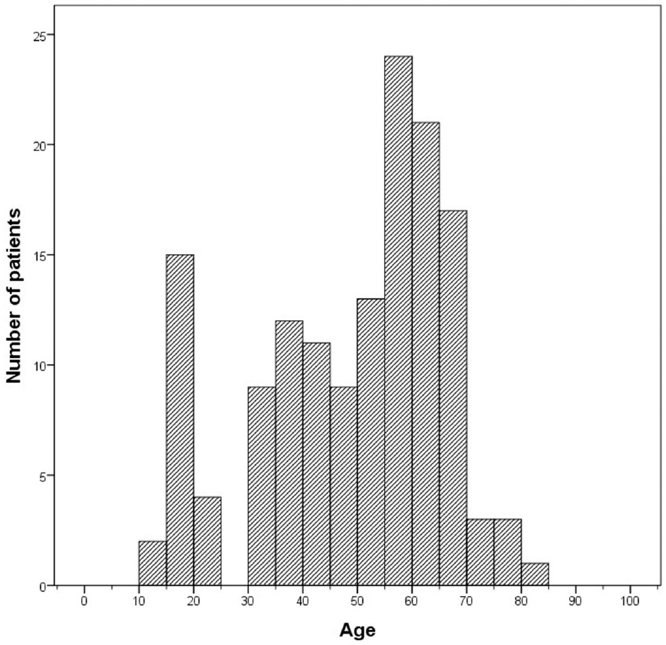
Figure 1.
Age-distribution of patients who underwent surgery for rhegmatogenous retinal detachment. The histogram shows bimodal distribution of rhegmatogenous retinal detachment by age.
 XML Download