

 PDF
PDF ePub
ePub Citation
 Print
Print


Abstract
Purpose
Methods
Results
Conclusions
Figures and Tables
Figure 1
Ultra-wide-field fundus photographs and ultra-wide-field fluorescein angiographic imaging of ocular toxocariasis. (A) A granuloma with mild vitreous opacity. (B) A tractional retinal fold with localized tractional retinal detachment. (C) Diffuse peripheral vascular leakage. (D) A prominent optic disc leakage.
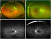
Figure 2
Ultra-wide-field fundus images with overlay of the Early Treatment Diabetic Retinopathy Study (ETDRS) 7-standard 30-degree fields. Small peripheral granuloma in nasal retina (A) and two peripheral granulomas in inferior retina (B) were noted in the image beyond the ETDRS 7 fields.
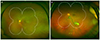
Figure 3
Ultra-wide-field fundus photographic findings of a patient with ocular toxocariasis before and after treatment of oral prednisolone with albendazole. (A) Severe vitreous opacity and multiple vitreous debris are shown. (B) The vitreous opacity was cleared up after treatment.
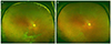
Figure 4
Ultra-wide-field fundus photographs of discontinuous migration of granuloma. (A) A round shape granuloma (white arrow) is shown. (B) After 3 months, a new granuloma (white arrow) appeared adjacent to the site of previous granuloma. (C) 1 week later, another novel granuloma (white arrow) was noted. (D) After 3 weeks, the granulomas were decreased in size.
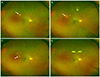
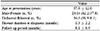
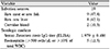
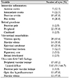
Table 4
Treatment outcomes in patients with ocular toxocariasis
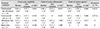
A p-value < 0.05 was considered statically significant.
N/A = not applicable; logMAR = logarithm of the minimum angle of resolution.
*p-values were obtained using the Wilcoxon singled-rank test; †Standardization of uveitis nomenclature (SUN) working group's grading scheme, based on the number of inflammatory cells present in a 1 × 1 mm slit beam of maximal intensity (grade 0: <1 cell, grade 0.5+: 1-5 cells, grade 1+: 6-15 cells, grade 2+: 16-25 cells, grade 4+: >50 cells); ‡National institutes of health (NIH) grading system for vitreous haze (grade 0: no flare, grade 0.5+: flare trace, grade 1+: clear optic disc and vessels, haze nerve fiber layer, grade 2+: hazy optic disc and vessels, grade 3+: optic disc visible, grade 4+: optic disc not visible).
 XML Download