

 PDF
PDF ePub
ePub Citation
 Print
Print


Abstract
Purpose
To evaluate the clinical efficacy of lacrimal endoscopy in patients with nasolacrimal duct obstruction (NLDO) and to compare the dacryocystography (DCG) and lacrimal endoscopic findings between patients with epiphora.
Methods
We conducted a retrospective chart review of 31 eyes of 23 patients who underwent an irrigation test, DCG, and lacrimal endoscopy from December 2014 to February 2016. We compared the clinical characteristics, and dacryocystographic findings, and lacrimal endoscopic findings of the patients, and analyzed whether or not these findings agree.
Results
Thirty-one eyes showed complete obstruction (13 eyes, 41.9%), partial obstruction (7 eyes, 22.6%), or patency (11 eyes, 35.5%) on irrigation test. Thirteen eyes with complete obstruction on irrigation test presented with complete obstruction (11 eyes, 84.6%) or secondary dilation (2 eyes, 15.4%) of the lacrimal sac at DCG. In terms of the level of obstruction, there was no difference between the two examinations. However, twelve eyes with complete obstruction at DCG; showed narrowing (4 eyes, 33%), granulation tissue (3 eye, 25%), mucus occlusion (2 eyes, 17%), stones (1 eye, 8%), or mucosal edema (2 eyes, 17%) on lacrimal endoscopy. Nineteen eyes with partial obstruction at DCG showed narrowing (6 eyes, 32%), mucus (5 eye, 26%), granulation tissue (4 eyes, 21%), or stones (4 eyes, 21%) on lacrimal endoscopy.
Conclusions
Lacrimal endoscopy allowed real-time observation inside the lacrimal passage that cannot be detected using DCG. Both methods provide comprehensive investigations of the nasolacrimal passage system, and these methods are complementary to understand the pathophysiology of nasolacrimal duct obstruction as well as planning treatment. Lacrimal endoscopy is very useful in investigating the lacrimal drainage passage in patients with NLDO, and this method is comparable to DCG.
Figures and Tables
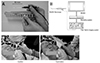 | Figure 1Lacrimal endoscopy (RUIDO fiberscope, Fibertechco., Tokyo, Japan). (A) Probe, Bent type, 0.9 mm diameter. (B) Peripheral device: the fibersope, monitor, recording system, imaging system. (C) The feature that applied to patient.
|
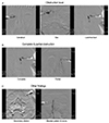 | Figure 2Representative figures of dacryocystography. (A) Images according to obstruction level; canalicular, sac, lacrimal duct. (B) Images according to the type of obstruction; complete, partial. (C) Other findings of lacrimal sac and nasolacrimal duct; secondary dilation, beaded pattern.
|
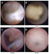 | Figure 3Representative photographs of lacrimal endoscopy. (A) The mucus occluding the lumen of the lacrimal canaliculus. (B) The stone inside of the lacrimal sac. (C) The granulation tissue occuping the lacrimal sac. (D) The stenosis of the nasolacrimal duct.
|
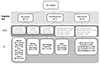 | Figure 4Correlation of dacryocystography (DCG) and lacrimal endoscopy (LE) findings based on the irrigation test in patients with nasolacrimal duct obstruction. The patients were divided into three groups according to the results of the syringing test, and categorized by DCG findings and lacrimal endoscopy findings.
|
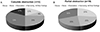 | Figure 5The result of lacrimal endoscopic findings according to the dacryocystgraphic finding in nasolacrimal duct obstruction patients. (A) 12 eyes with complete obstruction at dacryocystography (DCG); mucus (2 eyes, 17%), stone (1 eyes, 8%), granulation (3 eyes, 25%), narrowing (4 eyes, 33%). (B) 19 eyes with partial obstruction at DCG; mucus (5 eyes, 27%), stone (4 eyes, 21%), granulation (4 eyes, 21%), narrowing (6 eyes, 31%).
|
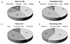 | Figure 6Result of dacryocystographic findings according to the lacrimal endoscopic finding in nasolacrimal duct obstruction patients. (A) 19 eyes with mucus at lacrimal endoscopy; narrowing (5 eyes, 26%), stone (3 eye, 16%), beaded pattern (3 eyes, 16%). (B) 14 eyes with granulation at lacrimal endoscopy; narrowing (6 eyes, 43%), no specific findings (6 eye, 43%), stone (1 eyes, 7%), beaded pattern (1 eyes, 7%). (C) 7 eyes with stone at lacrimal endoscopy; stone (3 eyes, 43%), no specific findings (3 eye, 43%), narrowing (1 eyes, 14%). (D) 21 eyes with narrowing at lacrimal endoscopy; narrowing (12 eyes, 57%), no specific findings (8 eye, 38%), beaded pattern (1 eyes, 4%).
|
References
1. Jones LT. An anatomical approach to problems of the eyelids and lacrimal apparatus. Arch Ophthalmol. 1961; 66:111–124.
2. Kanski JJ. of the lacrimal drainage system. Clinical Ophthalmology. Oxford: Butterworth-Heinemann;1997. p. 43–52.
3. Jeffrey JH, Myron Y, Jay SD. The lacrimal drainage system. Ophthalmology. 1999; 7:71–78.
4. Cohen SW, Prescott R, Sherman M, et al. Dacryoscopy. Ophthalmic Surg. 1979; 10:57–63.
5. Paulsen FP, Thale AB, Hallmann UJ, et al. The cavernous body of the human efferent tear ducts: function in tear outflow mechanism. Invest Ophthalmol Vis Sci. 2000; 41:965–970.
6. Paulsen FP, Corfield AP, Hinz M, et al. Characterization of mucins in human lacrimal sac and nasolacrimal duct. Invest Ophthalmol Vis Sci. 2003; 44:1807–1813.
7. Knop E, Knop N. Lacrimal drainage-associated lymphoid tissue (LDALT): a part of the human mucosal immune system. Invest Ophthalmol Vis Sci. 2001; 42:566–574.
8. Paulsen F, Thale A, Kohla G, et al. Functional anatomy of human lacrimal duct epithelium. Anat Embryol (Berl). 1998; 198:1–12.
9. Sasaki T, Nagata Y, Sugiyama K. Nasolacrimal duct obstruction classified by dacryoendoscopy and treated with inferior meatal dacryorhinotomy. Part I: Positional diagnosis of primary nasolacrimal duct obstruction with dacryoendoscope. Am J Ophthalmol. 2005; 140:1065–1069.
10. Sasaki T, Nagata Y, Sugiyama K. Nasolacrimal duct obstruction classified by dacryoendoscopy and treated with inferior meatal dacryorhinotomy: Part II. Inferior meatal dacryorhinotomy. Am J Ophthalmol. 2005; 140:1070–1074.
11. Sasaki H, Takano T, Murakami A. Direct endoscopic probing for congenital lacrimal duct obstruction. Clin Exp Ophthalmol. 2013; 41:729–734.
12. Maruyama N, Katori N, Sumiya N. Intraoperative use of the lacrimal endoscope for accurate reduction of bony nasolacrimal duct fracture. Plast Reconstr Surg. 2012; 130:761e–762e.
13. Kim CH, Lew H, Yun YS. Correspondence among the canaliculus irrigation test, dacryocystography and Jones test in the epiphora patients. J Korean Ophthalmol Soc. 2007; 48:1017–1022.
 XML Download