Abstract
Purpose
To investigate circumpapillary retinal nerve fiber layer (RNFL) and macular ganglion cell-inner plexiform layer (GCIPL) thicknesses as measured by optical coherence tomography in eyes with situs inversus of optic discs.
Methods
RNFL and macular GCIPL thicknesses were measured in eyes with situs inversus of optic discs without other ocular abnormalities (situs inversus group) and in age- and refractive error-matched healthy eyes (control group). RNFL thickness (global area, superior, nasal, inferior, and temporal quadrants) and GCIPL thickness (global area, superior-temporal, superior, superior-nasal, inferior-nasal, inferior, and inferior-temporal sectors and minimum) were compared between the groups.
Results
Nine eyes of 5 subjects with situs inversus of optic discs and 20 healthy eyes of 20 subjects (10 eyes for control groups A and B, respectively) were enrolled. No significant difference was found in superior or inferior quadrant RNFL thickness (p > 0.05); however, the situs inversus group showed a thicker RNFL in the nasal quadrant and a thinner RNFL in the temporal quad-rant (p < 0.01). In macular GCIPL thickness, no significant difference was found in the superotemporal or inferiotemporal sector or for minimum thickness (p < 0.05); however, the situs inversus group showed thicker GCIPL in the global area, superior, super-onasal, inferonasal, and inferior sectors than the control groups (p < 0.01).
Go to : 
References
3. Kothari M, Chatterjee DN. . Unilateral situs inversus of optic disc as-sociated with reduced binocularity and stereoacuity resembling monofixation syndrome. Indian J Ophthalmol. 2010; 58:241–2.

4. Kang S, Jin S, Roh KH, Hwang YH. . Peripapillary retinal nerve fi-ber layer and optic nerve head characteristics in eyes with situs in-versus of the optic disc. J Glaucoma. 2015; 24:306–10.

5. Han SY, Hwang YH. . Glaucoma in an eye with situs inversus of the optic disc. Semin Ophthalmol. 2014; 29:172–4.

6. Mwanza JC, Durbin MK, Budenz DL. . Glaucoma diagnostic accuracy of ganglion cell-inner plexiform layer thickness: compar-ison with nerve fiber layer and optic nerve head. Ophthalmology. 2012; 119:1151–8.

7. Jeoung JW, Choi YJ, Park KH, Kim DM. . Macular ganglion cell imaging study: glaucoma diagnostic accuracy of spectral-domain optical coherence tomography. Invest Ophthalmol Vis Sci. 2013; 54:4422–9.

Go to : 
 | Figure 1.An example of optic disc in a healthy eye (left col-umn) and an eye with situs inversus of optic disc (right col-umn). Disc photograph (A), red-fee fundus photograph (B), and circumpapillary retinal nerve fiber layer thickness dis-tribution as measured by Cirrus high– definition optical coher-ence tomography(C). |
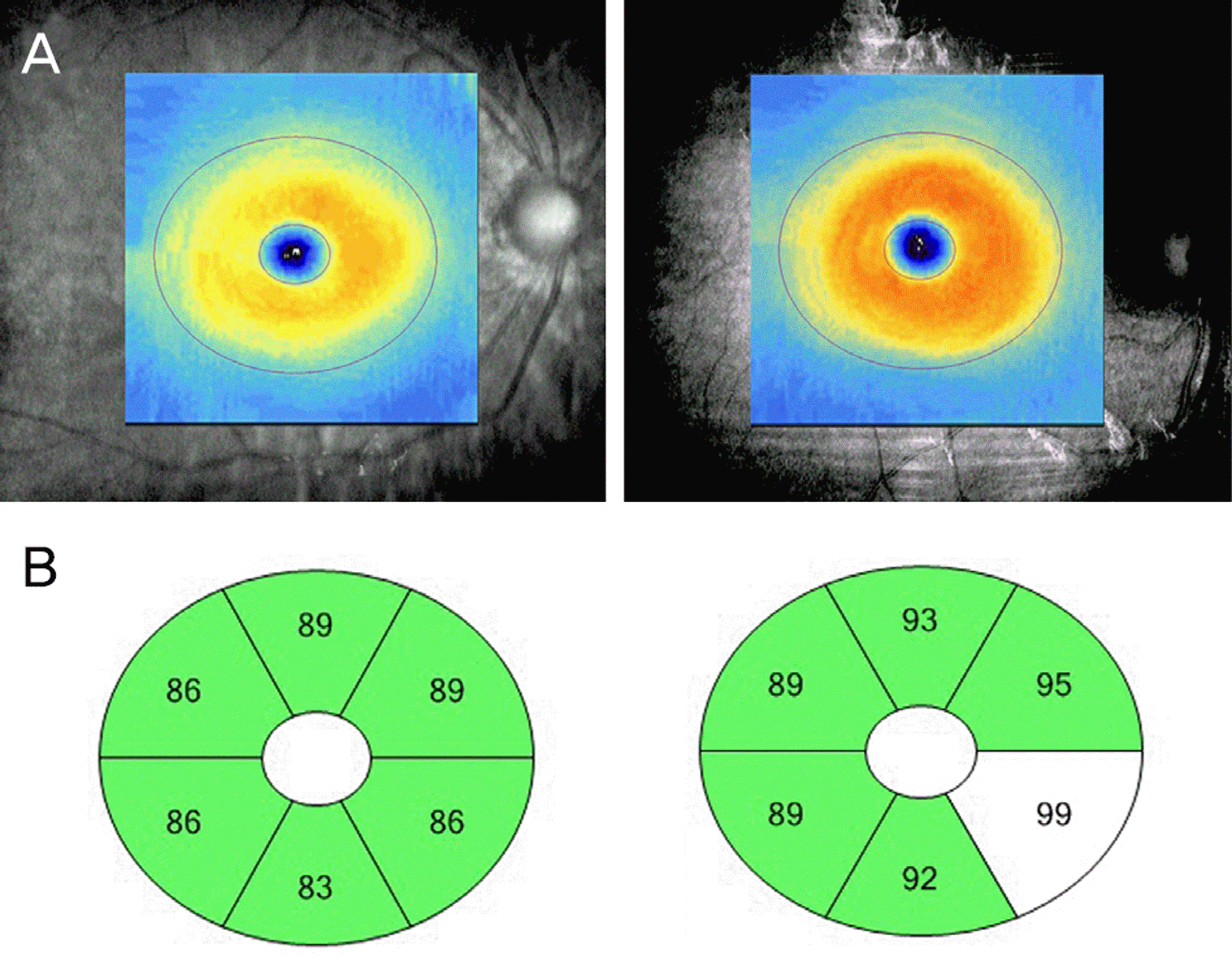
 | Figure 2.An example of macular ganglion cell-inner plexi-form layer thickness distribution. Presented by thickness map (A) and sector map (B) of Cirrus high– definition optical co-herence tomography in a healthy eye (left column) and an eye with situs inversus of optic disc (right column) presented in Figure 1. |
Table 1.
Comparison of clinical data between control and situs inversus groups
| Control group A (n = 10) | Control group B (n = 10) | Situs inversus group (n = 9) | p-value* | p-value† | |
|---|---|---|---|---|---|
| Age (years) | 43.0 (39.0, 46.3) | 44.5 (29.0, 52.0) | 48.0 (33.0, 49.0) | 0.905 | 0.842 |
| Sex (female:male) | 5:5 | 5:5 | 4:6 | 1.000 | 1.000 |
| Refractive error (diopter) | -0.63 (-1.00, -0.38) | -1.00 (-1.57, -0.69) | -1.00 (-2.00, -0.25) | 0.315 | 0.720 |
| Visual field indices | |||||
| Mean deviation (dB) | -0.79 (-1.89, 0.30) | -1.19 (-1.68, -0.08) | -0.85 (-1.28, 0.72) | 0.497 | 0.182 |
| Pattern standard deviation (dB) | 1.55 (1.48, 1.94) | 1.44 (1.14, 1.89) | 1.67 (1.30, 2.21) | 0.661 | 0.243 |
| Visual field index (%) | 99.0 (99.0, 99.25) | 99.5 (99.0, 100.0) | 100.0 (98.5, 100.0) | 0.400 | 0.968 |
Table 2.
Comparison of circumpapillary retinal nerve fiber layer thickness (μ m) between control and situs inversus groups
| Control group A (n = 10) | Control group B (n = 10) | Situs inversus group (n = 9) | p-value* | p-value† | |
|---|---|---|---|---|---|
| Average | 95.5 (87.5, 99.5) | 100.0 (95.8, 108.3) | 102.0 (94.5, 105.5) | 0.211 | 0.905 |
| Superior quadrant | 124.0 (114.5, 143.3) | 130.0 (119.8, 134.5) | 118.0 (111.0, 143.0) | 0.720 | 0.604 |
| Nasal quadrant | 60.0 (51.5, 70.3) | 64.5 (62.5, 76.3) | 82.0 (71.5, 105.0) | 0.004 | 0.010 |
| Inferior quadrant | 122.0 (110.0, 132.5) | 130.5 (125.5, 142.5) | 126.0 (123.0, 146.0) | 0.211 | 0.661 |
| Temporal quadrant | 70.5 (61.0, 77.0) | 79.0 (73.0, 84.5) | 56.0 (52.0, 60.5) | 0.001 | <0.001 |
| Clock-hour sectors | |||||
| 12 | 135.5 (120.0, 155.0) | 122.5 (102.8, 137.8) | 131.0 (118.5, 156.5) | 0.661 | 0.400 |
| 1 | 112.5 (104.8, 122.0) | 111.5 (94.8, 114.0) | 146.0 (101.5, 188.0) | 0.133 | 0.065 |
| 2 | 68.0 (58.3, 82.3) | 75.0 (68.5, 88.0) | 99.0 (81.0, 132.5) | 0.006 | 0.013 |
| 3 | 52.5 (43.5, 61.0) | 59.5 (55.0, 68.0) | 76.0 (57.5, 88.0) | 0.022 | 0.156 |
| 4 | 65.0 (52.0, 66.8) | 62.5 (57.3, 73.3) | 92.0 (70.5, 103.0) | 0.002 | 0.017 |
| 5 | 89.5 (85.8, 109.5) | 88.5 (84.8, 106.5) | 141.0 (115.0, 173.0) | 0.002 | 0.002 |
| 6 | 137.0 (108.0, 163.8) | 128.5 (123.8, 161.5) | 162.0 (156.5, 167.0) | 0.095 | 0.043 |
| 7 | 135.0 (130.8, 156.5) | 165.0 (149.5, 173.5) | 90.0 (80.0, 101.5) | <0.001 | <0.001 |
| 8 | 70.0 (62.8, 80.8) | 79.5 (72.0, 92.3) | 54.0 (41.5, 58.0) | 0.001 | <0.001 |
| 9 | 57.0 (51.0, 62.3) | 57.5 (51.3, 70.0) | 52.0 (47.0, 58.0) | 0.133 | 0.133 |
| 10 | 75.5 (68.8, 89.0) | 93.0 (89.5, 103.5) | 66.0 (60.5, 71.0) | 0.022 | <0.001 |
| 11 | 120.0 (114.8, 133.0) | 151.5 (139.0, 168.0) | 89.0 (77.5, 112.5) | 0.002 | <0.001 |
Table 3.
Comparison of macular ganglion cell-inner plexiform layer thickness (μ m) between control and situs inversus groups
| Control group A (n = 10) | Control group B (n = 10) | Situs inversus group (n = 9) | p-value* | p-value† | |
|---|---|---|---|---|---|
| Average | 82.0 (81.0, 87.0) | 84.5 (81.8, 86.0) | 90.0 (83.5, 91.5) | 0.035 | 0.028 |
| Minimum | 81.5 (79.8, 83.8) | 82.0 (80.0, 85.0) | 84.0 (80.5, 87.0) | 0.315 | 0.447 |
| Superotemporal | 82.5 (79.8, 85.3) | 84.0 (79.8, 86.0) | 83.0 (81.5, 88.0) | 0.356 | 0.549 |
| Superior | 82.5 (81.5, 85.8) | 84.0 (82.3, 85.3) | 89.0 (86.0, 93.0) | 0.010 | 0.008 |
| Superonasal | 85.0 (83.8, 88.5) | 87.0 (84.75, 88.0) | 94.0 (88.5, 95.5) | 0.004 | 0.013 |
| Inferonasal | 83.5 (81.8, 87.3) | 85.5 (82.3, 86.0) | 93.0 (85.0, 96.5) | 0.028 | 0.017 |
| Inferior | 80.0 (78.8, 85.3) | 82.0 (79.5, 85.3) | 89.0 (81.0, 91.5) | 0.033 | 0.059 |
| Inferotemporal | 81.5 (79.0, 86.5) | 83.5 (81.5, 86.3) | 86.0 (80.0, 88.5) | 0.315 | 0.661 |


 PDF
PDF ePub
ePub Citation
 Print
Print


 XML Download