Journal List > J Korean Ophthalmol Soc > v.58(4) > 1010740

Lee, Kim, Kim, Shin, Kim, Lee, and Lee: Prognostic Factors of Neovascular Glaucoma in Eyes with Treated Proliferative Diabetic Retinopathy

Abstract

Purpose

To evaluate the factors that are significant in progression to neovascular glaucoma in patients with proliferative diabetic retinopathy.

Methods

In this retrospective analysis, we reviewed the medical records of 52 patients who were first diagnosed with proliferative diabetic retinopathy from March 2014 to March 2016. We compared diabetes mellitus period, HbA1c, chronic diseases such as hypertension, hyperlipidemia, and kidney disease, insulin treatments, blood urea nitrogen, blood creatinine, glomerular filtration rate, urine albumin, dialysis, corrected visual acuity at the first visit, traction membrane sign of the retina at the first visit, vitreous hemorrhage and preretinal hemorrhage in each group and then investigated the prognostic factors of neovascular glaucoma.

Results

A total of 52 patients were included in the investigation, 12 patients (23.08%) were diagnosed with iris neovascularization and 4 patients (7.69%) developed neovascular glaucoma. The patients without iris neovascularization were defined as Group I, those with iris neovascularization as Group II, and those with neovascular glaucoma as Group III. The diabetes mellitus period was significantly longer in Group II (10.88 ± 7.14 years) and in Group III (11.75 ± 8.61 years) than Group I (8.30 ± 5.25 years) (p-value 0.41, 0.032, respectively). The HbA1c level was 9.59 ± 2.23 in Group II and 9.27 ± 2.54 in Group I. There was no significant difference between the two groups (p = 0.721). However, HbA1c was significantly higher in Group III (11.55 ± 0.21) than Group I (p-value 0.048).

Conclusions

A long diabetes mellitus period and high HbA1c level have a significant effect on the progression to neovascular glaucoma in patients with proliferative diabetic retinopathy. This information could be useful for predicting and preventing the prognosis of patients.

Figures and Tables

Table 1

Demographics of three groups

jkos-58-415-i001

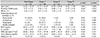
Values are presented as mean ± SD unless otherwise indicated. ‘Group I’ means ‘Control Group’, ‘Group II’ means ‘patients who developed neovascular iridis’, and ‘Group III’ means ‘Patients who developed neovascular glaucoma’.

SD = standard deviation; DM = diabetic mellitus; BUN = blood urea nitrogen; Cr = Creatinine; GFR = glomerular filtration rate.

*Statistically significant; Comparison between Group I and Group II (Student t-test for continuous variables, chai-square test for discontinuous variables); Comparison between Group I and Group III (Mann-Whitney U-test for continuous variables, chai-square test for discontinuous variables).

Table 2

Demographics of three groups according to ophthalmologic examination

jkos-58-415-i002

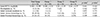
Values are presented as mean ± SD or n (%) unless otherwise indicated. ‘Group I’ means ‘Control Group’, ‘Group II’ means ‘Patients who developed neovascular iridis’ and ‘Group III’ means ‘Patients who developed neovascular glaucoma’.

SD = standard deviation; BCVA = best corrected visual acuity.

*Comparison between Group I and Group II (Student t-test for continuous variables, chai-square test for discontinuous variables); Comparison between Group I and Group III (Mann-Whitney U-test for continuous variables, chai-square test for discontinuous variables).

References

1. Sivak-Callcott JA, O'Day DM, Gass JD, Tsai JC. Evidence-based recommendations for the diagnosis and treatment of neovascular glaucoma. Ophthalmology. 2001; 108:1767–1776. quiz 1777, 1800.
2. Ohrt V. The frequency of rubeosis iridis in diabetic patients. Acta Ophthalmol (Copenh). 1971; 49:301–307.
3. Madsen PH. Rubeosis of the iris and haemorrhagic glaucoma in patients with proliferative diabetic retinopathy. Br J Ophthmol. 1971; 55:368–371.
4. Shazly TA, Latina MA. Neovascular glaucoma: etiology, diagnosis and prognosis. Semin Ophthalmol. 2009; 24:113–121.
5. Klein R, Klein BE, Moss SE, et al. The Wisconsin Epidemiologic study of diabetic retinopathy. IV. Diabetic macular edema. Ophthalmology. 1984; 91:1464–1474.
6. Klein R, Klein BE, Moss SE, Cruickshanks KJ. The Wisconsin epidemiologic study of diabetic retinopathy. XV. The long-term incidence of macular edema. Ophthalmology. 1995; 102:7–16.
7. Tripathi RC, Li J, Tripathi BJ, et al. Increased level of vascular endothelial growth factor in queous humor of patients with neovascular glaucoma. Ophthalmology. 1998; 105:232–237.
8. Wand M, Madigan JC, Gaudio AR, Sorokanich S. Neovascular glaucoma following pars plana vitrectomy for complication of diabetic retinopathy. Ophthalmic Surg. 1990; 21:113–118.
TOOLS
Similar articles