Abstract
Purpose
To evaluate the long-term changes in cornea endothelial cell density (ECD) after pars plana vitrectomy (PPV) with fragmentation.
Methods
Twenty patients (20 eyes) who underwent PPV with fragmentation and who were followed up for 2 years were enrolled in this retrospective study. The cornea ECD, coefficient of variation (CV), and hexagonality (HA) were calculated using a spec-ular microscopy at 1, 3, 6, 12 months, and 2 years after surgery.
Results
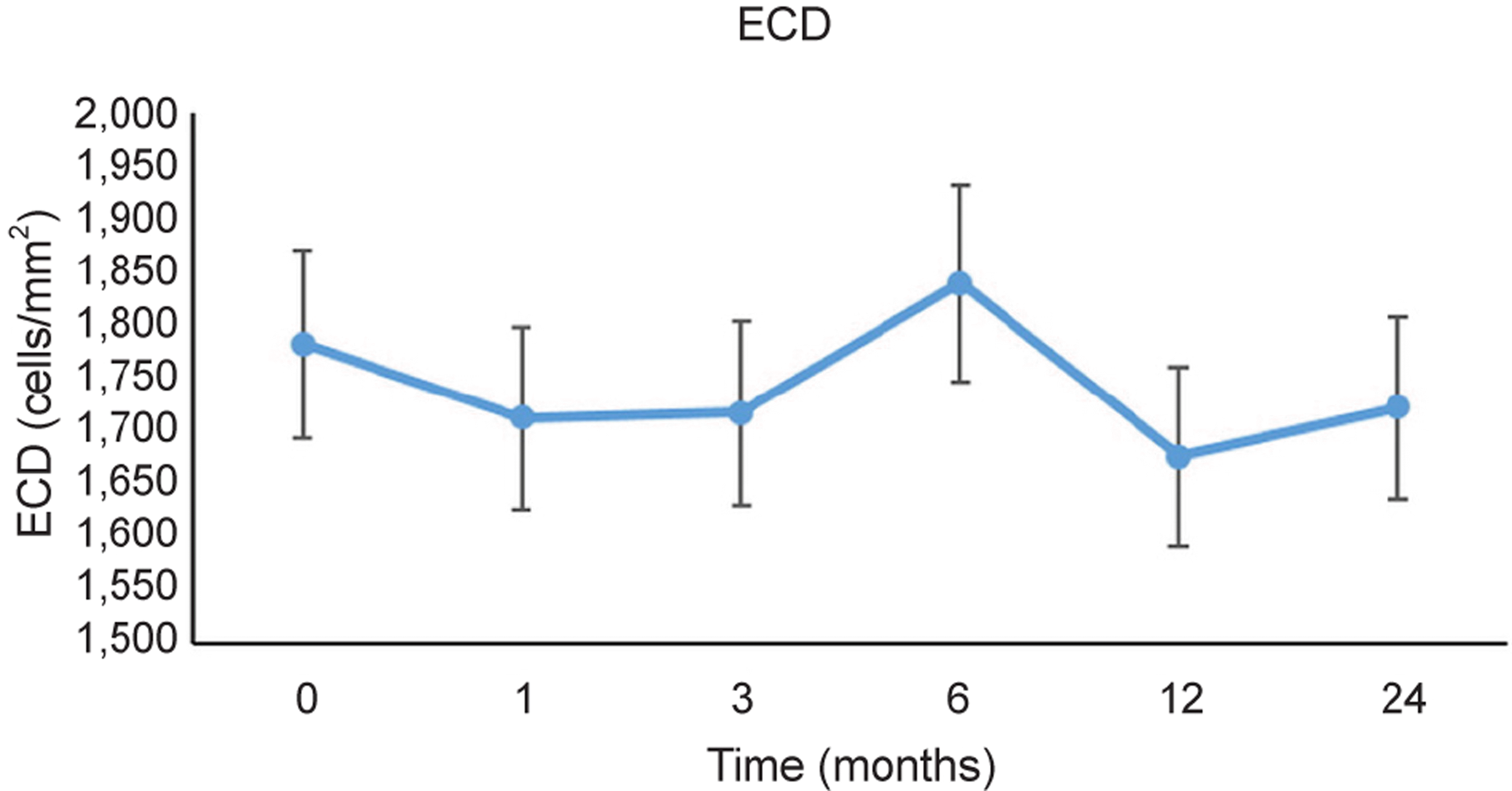
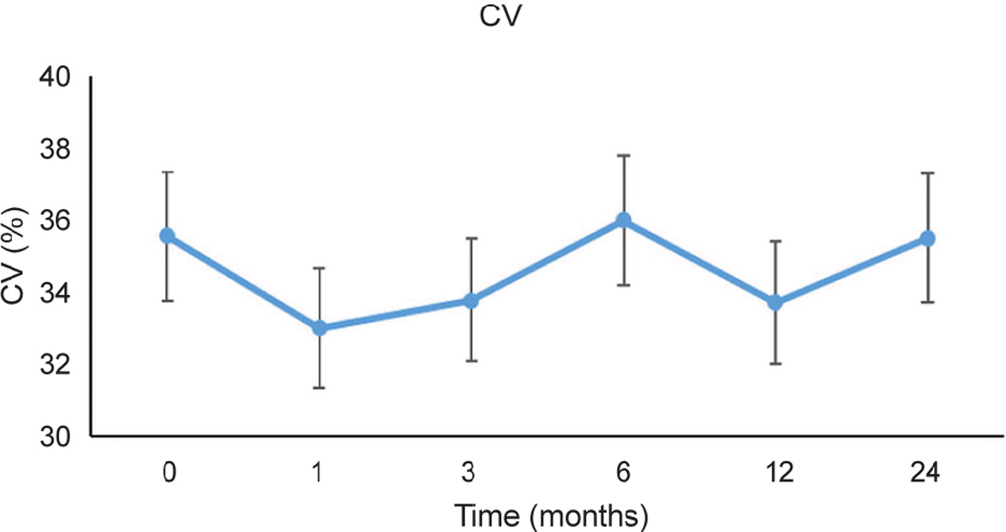
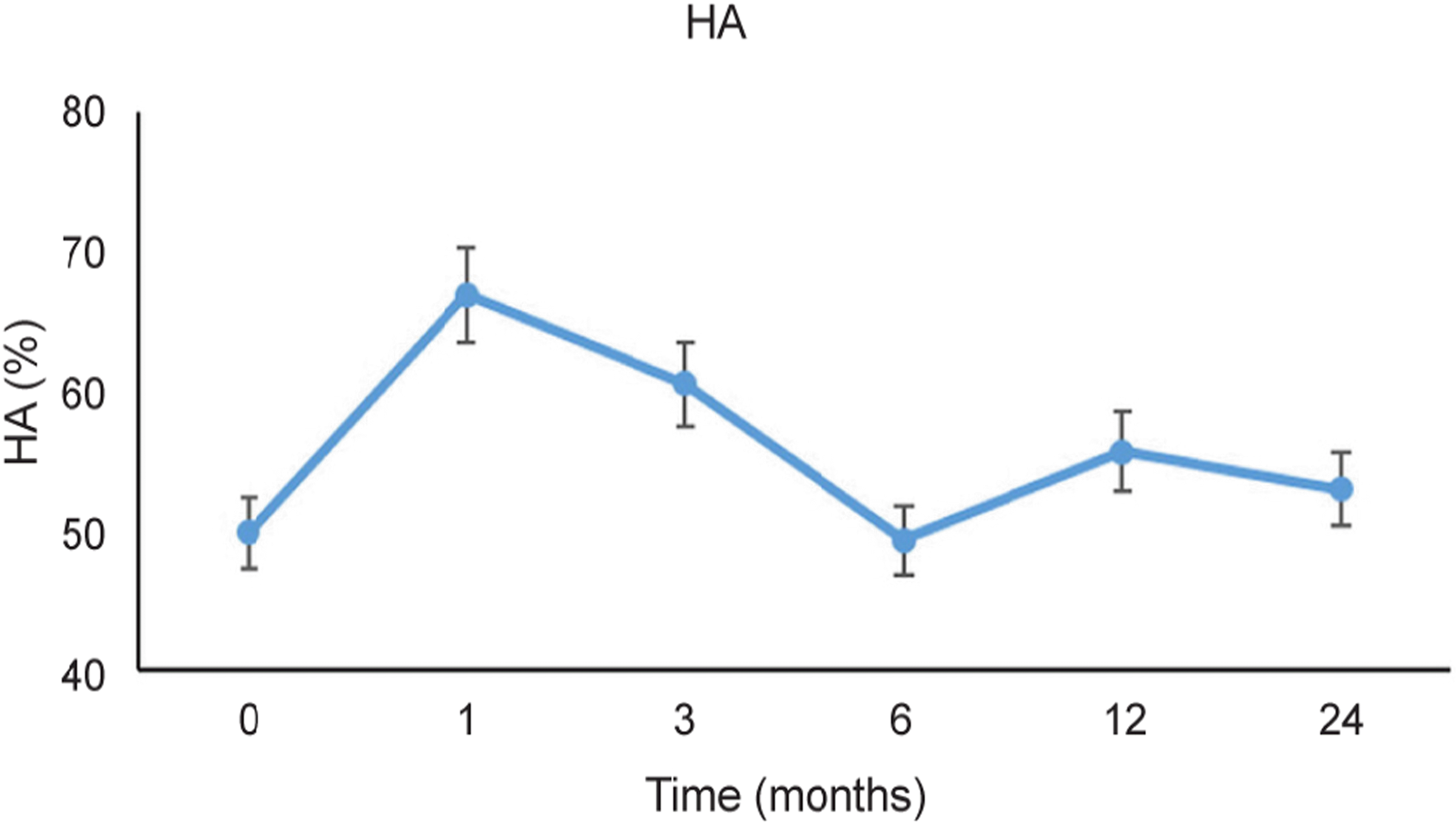
The preoperative mean ECD was 1,782 ± 623 cells/mm2, and the postoperative mean ECD did not significantly change at 1, 3, 6, and 12 months. Additionally, there were no significant changes in CV or HA. At 2 years after surgery, the mean ECD was 1,722 ± 532 cells/mm2, the mean CV was 35.50 ± 3.03%, and the mean HA was 53.00 ± 4.91%. There were no significant changes in ECD, CV, or HA preoperatively and postoperatively at 1, 3, 6, and 12 months.
Go to : 
References
1. Waring GO 3rd, Bourne WM, Edelhauser HF, Kenyon KR. . The corneal endothelium. Normal and pathologic structure and function. Ophthalmology. 1982; 89:531–90.

2. Joyce NC. . Proliferative capacity of the corneal endothelium. Prog Retin Eye Res. 2003; 22:359–89.

3. Werblin TP. . Long-term endothelial cell loss following phacoe-mulsification: model for evaluating endothelial damage after intra-ocular surgery. Refract Corneal Surg. 1993; 9:29–35.

4. Baradaran-Rafii A, Rahmati-Kamel M, Eslani M. . Effect of hydrodynamic parameters on corneal endothelial cell loss after phacoemulsification. J Cataract Refract Surg. 2009; 35:732–7.

5. Bourne RR, Minassian DC, Dart JK. . Effect of cataract sur-gery on the corneal endothelium: modern phacoemulsification compared with extracapsular cataract surgery. Ophthalmology. 2004; 111:679–85.
6. Faramarzi A, Javadi MA, Karimian F. . Corneal endothelial cell loss during phacoemulsification: bevel-up versus bevel-down phaco tip. J Cataract Refract Surg. 2011; 37:1971–6.

7. Diddie KR, Schanzlin DJ. . Specular microscopy in pars plana vitrectomy. Arch Ophthalmol. 1983; 101:408–9.

8. Friberg TR, Doran DL, Lazenby FL. . The effect of vitreous and reti-nal surgery on corneal endothelial cell density. Ophthalmology. 1984; 91:1166–9.

9. Koenig SB, Mieler WF, Han DP, Abrams GW. . Combined phacoe-mulsification, pars plana vitrectomy, and posterior chamber intra-ocular lens insertion. Arch Ophthalmol. 1992; 110:1101–4.

10. Yeniad B, Corum I, Ozgun C. . The effects of blunt trauma and cata-ract surgery on corneal endothelial cell density. Middle East Afr J Ophthalmol. 2010; 17:354–8.

11. Eom Y, Kim SW, Ahn J. . Comparison of cornea endothelial cell counts after combined phacovitrectomy versus pars plana vi-trectomy sith fragmentation. Graefes Arch Clin Exp Ophthalmol. 2013; 251:2187–93.
12. Kim JT, Eom Y, Ahn J. . The use of a 20-gauge valved cannula during pars plana phacofragmentation with a 23-gauge ultrasonic fragmatome. Ophthalmic Surg Lasers Imaging Retina. 2014; 45:207–10.

13. Seki M, Yammamoto S, Abe H, Fukuchi T. . Modified ab externo method for introducing 2 polypropylene loops for scleral suture fixation of intraocular lenses. J cataract Refract Surg. 2013; 39:1291–6.

14. Heuermann T, Hartmann C, Anders N. . Long-term endothelial cell loss after phacoemulsification: peribulbar anesthesia versus intra-cameral lidocaine 1%. prospective randomized clinical trial. J Cataract Refract Surg. 2002; 28:639–43.

15. Park HY, Lee NY, Park CK, Kim MS. . Long-term changes in endo-thelial cell counts after early phacoemulsification versus laser pe-ripheral iridotomy using sequential argon: YAG laser technique in acute primary angle closure. Graefes Arch Clin Exp Ophthalmol. 2012; 250:1673–80.
16. Linebarger EJ, Hardten DR, Shah GK, Lindstrom RL. . Phacoemulsi- fication and modern cataract surgery. Surv Ophthalmol. 1999; 44:123–47.
17. Kim EK, Cristol SM, Geroski DH. . Corneal endothelial dam-age by air bubbles during phacoemulsification. Arch Ophthalmol. 1997; 115:81–8.

18. Cinar E, Zengin MO, Kucukerdonmez C. . Evaluation of corneal en-dothelial cell damage after vitreoretinal surgery: comparison of different endotamponades. Eye (Lond). 2015; 29:670–4.

19. Farrahi F, Feghhi M, Ostadian F, Alivand A. . Pars plana vitrectomy and silicone oil injection in phakic and pseudophakic eyes; corneal endothelial changes. J Ophthalmic Vis Res. 2014; 9:310–3.
20. Mitamura Y, Yamamoto S, Yamazaki S. . Corneal endothelial cell loss in eyes undergoing lensectomy with and without anterior lens capsule removal combined with pars plana vitrectomy and gas tamponade. Retina. 2000; 20:59–62.

21. Mittl RN, Koester CJ, Kates MR, Wilkes E. . Endothelial cell counts following pars plana vitrectomy in pseudophakic and aphakic eyes. Ophthalmic Surg. 1989; 20:13–6.

22. Nadal J, Kudsieh B, Casaroli-Marano RP. . Scleral fixation of poste-riorly dislocated intraocular lenses by 23-gauge vitrectomy with-out anterior segment approach. J Ophthalmol. 2015; 2015:391619.

23. Mutoh T, Matsumoto Y, Chikuda M. . Scleral fixation of foldable acrylic intraocular lenses in aphakic post-vitrectomy eyes. Clin Ophthalmol. 2010; 5:17–21.

24. Nishida T, Saika S. . Cornea and sclera: anatomy and physiology. Krachmer JH, Mannis MJ, Holland EJ, editors. Cornea. 3rd ed.St. Louis: Mosby;2011; chap. 1.
25. Kim KS, Park SY, Oh JS. . Morphometric analysis of the corneal en-dothelial cells in normal Korean. J Korean Ophthalmol Soc. 1992; 33:320–5.
Go to : 
 | Figure 1.Changes in endothelial cell density(ECD). No sig-nificant changes in ECD at 1, 3, 6, 12 and 24 months after pars plana vitrectomy with fragmentation. |
 | Figure 2.Changes in coefficient of variation (CV) of endothe-lial cells. No significant changes in CV at 1, 3, 6, 12 and 24 months after pars plana vitrectomy with fragmentation. |
 | Figure 3.Changes in endothelial cell hexagonality (HA). No significant changes in HA at 1, 3, 6, 12 and 24 months after pars plana vitrectomy with fragmentation. |
Table 1.
Baseline clinical characteristics
Table 2.
Surgical indications for pars plana vitrectomy
| PPV with fragmentation (n = 20) | |
|---|---|
| Epiretinal membrane | 2 (10.0) |
| BRVO hemorrhage | 2 (10.0) |
| Low endothelial cell counts | 10 (50.0) |
| Zonulysis | 6 (30.0) |
Table 3.
Changes in endothelial cell density (ECD)
| Period | No. of Eyes | Mean ECD (cells/mm2) | Mean observed percentage ECD change | p-value* |
|---|---|---|---|---|
| Preoperative | 20 | 1,782 ± 623 | NA | NA |
| Postop 1 month | 19 | 1,665 ± 685 | 4.52 ± 16.78 | 0.953 |
| Postop 3 months | 13 | 1,791 ± 518 | -2.72 ± 21.48 | 0.980 |
| Postop 6 months | 13 | 1,859 ± 719 | 11.92 ± 39.48 | 0.989 |
| Postop 1 year | 17 | 1,642 ± 695 | 5.02 ± 19.85 | 0.808 |
| Postop 2 years | 20 | 1,722 ± 532 | -1.30 ± 26.94 | 0.973 |
Table 4.
Changes in coefficient of variation (CV)
| Period | No. of Eyes | Mean CV | Mean observed percentage CV change | p-value* |
|---|---|---|---|---|
| Preoperative | 20 | 35.55 ± 3.03 | NA | NA |
| Postop 1 month | 19 | 33.00 ± 3.06 | -3.31 ± 29.57 | 0.932 |
| Postop 3 months | 12 | 33.78 ± 3.46 | -2.54 ± 41.96 | 0.993 |
| Postop 6 months | 13 | 35.98 ± 3.38 | 9.01 ± 30.80 | 1.000 |
| Postop 12 months | 16 | 33.70 ± 3.19 | 0.35 ± 27.01 | 0.986 |
| Postop 2 years | 20 | 35.50 ± 3.03 | 3.45 ± 24.69 | 1.000 |
Table 5.
Changes in hexagonality (HA)
| Period | No. of Eyes | Mean HA (%) | Mean observed percentage HA change | p-value* |
|---|---|---|---|---|
| Preoperative | 15 | 49.86 ± 5.60 | NA | NA |
| Postop 1 month | 18 | 66.88 ± 5.15 | -77.44 ± 125.85 | 0.141 |
| Postop 3 months | 11 | 60.48 ± 6.74 | -48.44 ± 67.26 | 0.776 |
| Postop 6 months | 12 | 49.35 ± 6.19 | -49.61 ± 132.89 | 1.000 |
| Postop 12 months | 16 | 55.70 ± 5.43 | -75.79 ± 128.32 | 0.960 |
| Postop 2 years | 20 | 53.00 ± 4.91 | -38.08 ± 122.78 | 0.997 |
Table 6.
Comparison of ECD, CV and HA of corneal endothelial cells after pars plana vitrectomy with fragmentation
| Period | p-value* | ||
|---|---|---|---|
| ECD | CV | HA | |
| Baseline vs. postop 1 month | 0.953 | 0.932 | 0.141 |
| Baseline vs. postop 3 months | 0.980 | 0.993 | 0.776 |
| Baseline vs. postop 6 months | 0.989 | 1.000 | 1.000 |
| Baseline vs. postop 12 months | 0.808 | 0.986 | 0.960 |
| Baseline vs. postop 2 years | 0.973 | 1.000 | 0.997 |
| Postop 1 month vs. 3 months | 1.000 | 0.999 | 0.962 |
| Postop 1 month vs. 6 months | 0.746 | 0.927 | 0.180 |
| Postop 1 month vs. 12 months | 0.998 | 0.999 | 0.557 |
| Postop 1 month vs. 2 years | 1.000 | 0.937 | 0.252 |
| Postop 3 month vs. 6 months | 0.848 | 0.989 | 0.790 |
| Postop 3 month vs. 12 months | 0.998 | 1.000 | 0.991 |
| Postop 3 month vs. 2 years | 1.000 | 0.994 | 0.924 |
| Postop 6 month vs. 12 months | 0.540 | 0.980 | 0.959 |
| Postop 6 month vs. 2 years | 0.793 | 1.000 | 0.996 |
| Postop 1 year vs. 2 years | 0.994 | 0.988 | 0.998 |


 PDF
PDF ePub
ePub Citation
 Print
Print


 XML Download