Abstract
Purpose
To evaluate the short-term changes in tear film dynamics with non-invasive infrared imaging measurement before and after cataract surgery as a pilot study.
Methods
Seventeen eyes of 17 patients without preoperative dry eye were enrolled in this study. Non-invasive keratograph tear break-up time (NIK-TBUT) and non-invasive keratograph tear meniscus height (NIK-TMH) were measured before and 1 day, 1 week, and 1 month after cataract surgery using a keratograph.
Results
The mean patient age was 64.47 ± 10.28 years, and 78.95% were female. Although the mean postoperative 1 day NIK-TBUT-first value was not significantly different from the preoperative value, the mean postoperative 1 week and 1 month NIK-TBUT-first values were significantly lower than preoperative values (p < 0.05). The postoperative 1 day, 1 week, and 1 month NIK-TBUT-average and the NIK-TMH values were not different from preoperative values.
Conclusions
Our study showed that cataract surgery can lead to tear film instability. And it is important to determine the long-term outcomes of surgery and whether NIK-TBUT and NIK-TMH correlate with slit lamp examination tear break-up time (SLE-TBUT) and slit lamp examination tear meniscus height (SLE-TMH).
Go to : 
References
1. The definition and classification of dry eye disease: report of the definition and classification subcommittee of the international dry eye workshop . . Ocular surf. 2007; 5:75–92.
2. The epidemiology of dry eye disease: report of the epidemiology subcommittee of the international dry eye workshop . . Ocular surf. 2007; 5:93–107.
3. Li XM, Hu L, Hu J, Wang W. . Investigation of dry eye disease and analysis of the pathogenic factors in patients after cataract surgery. Cornea. 2007; 26:(9 Suppl 1):S16-20.

4. Cetinkaya S, Mestan E, Acir NO. . The course of dry eye after phacoemulsification surgery. BMC Ophthalmol. 2015; 15:68.

5. Cho YK, Kim MS. . Dry eye after cataract surgery and associated in-traoperative risk factors. Korean J Ophthalmol. 2009; 23:65–73.

6. Kasetsuwan N, Satitpitakul V, Changul T, Jariyakosol S. . Incidence and pattern of dry eye after cataract surgery. PLoS One. 2013; 8:e78657.

7. Cox SM, Nichols KK, Nichols JJ. . Agreement between automated and traditional measures of tear film breakup. Optom Vis Sci. 2015; 92:e257–63.

8. Lan W, Lin L, Yang X, Yu M. . Automatic noninvasive tear breakup time (TBUT) and conventional fluorescent TBUT. Optom Vis Sci. 2014; 91:1412–8.

9. Yokoi N, Komuro A. . Non-invasive methods of assessing the tear film. Exp Eye Res. 2004; 78:399–407.

10. Abdelfattah NS, Dastiridou A, Sadda SR, Lee OL. . Noninvasive imaging of tear film dynamics in eyes with ocular surface disease. Cornea. 2015; 34:Suppl 10:S48-52.

11. Nichols KK, Mitchell GL, Zadnik K. . The repeatability of clinical measurements of dry eye. Cornea. 2004; 23:272–85.

12. Dogru M, Ishida K, Matsumoto Y. . Strip meniscometry: a new and simple method of tear meniscus evaluation. Invest Ophthalmol Vis Sci. 2006; 47:1895–901.

13. Altan-Yaycioglu R, Sizmaz S, Canan H, Coban-Karatas M. . Optical coherence tomography for measuring the tear film meniscus: cor-relation with schirmer test and tear-film breakup time. Curr Eye Res. 2013; 38:736–42.

14. Madden RK, Paugh JR, Wang C. . Comparative study of two non-in-vasive tear film stability techniques. Curr Eye Res. 1994; 13:263–9.

15. Goto T, Zheng X, Okamoto S, Ohashi Y. . Tear film stability analysis system: introducing a new application for videokeratography. Cornea. 2004; 23:(8 Suppl):S65-70.
16. Jiang Y, Ye H, Xu J, Lu Y. . Noninvasive keratograph assessment of tear film break-up time and location in patients with age-related cataracts and dry eye syndrome. J Int Med Res. 2014; 42:494–502.

17. Hong J, Sun X, Wei A. . Assessment of tear film stability in dry eye with a newly developed keratograph. Cornea. 2013; 32:716–21.

18. Kohlhaas M. . Corneal sensation after cataract and refractive surgery. J Cataract Refract Surg. 1998; 24:1399–409.

19. Ram J, Gupta A, Brar G. . Outcomes of phacoemulsification in patients with dry eye. J Cataract Refract Surg. 2002; 28:1386–9.

20. Oh T, Jung Y, Chang D. . Changes in the tear film and ocular surface after cataract surgery. Jpn J Ophthalmol. 2012; 56:113–8.

21. Khanal S, Tomlinson A, Esakowitz L. . Changes in corneal sensitivity and tear physiology after phacoemulsification. Ophthalmic Physiol Opt. 2008; 28:127–34.

22. Methodologies to diagnose and monitor dry eye disease: report of the definition and classification subcommittee of the international dry eye workshop . . Ocular Surf. 2007; 5:108–52.
23. Kojima T, Ishida R, Dogru M. . A new noninvasive tear stabil-ity analysis system for the assessment of dry eyes. Invest Ophthalmol Vis Sci. 2004; 45:1369–74.

Go to : 
 | Figure 1.Screenshot of output window of Keratograph 5M for the left eye of a patient. Left panel: a dynamic video recording captures the process of tear break up. Upper right panel: the total break-up areas during the time when the eye was open. Lower right panel: In the patient, non-invasive Keratography break-up time first (NIKBUT-first) is 4.7 seconds, non-invasive Keratograph break-up time-average (NIKBUT-average) is 8.6 seconds and automatic dry eye classification is level 1. |
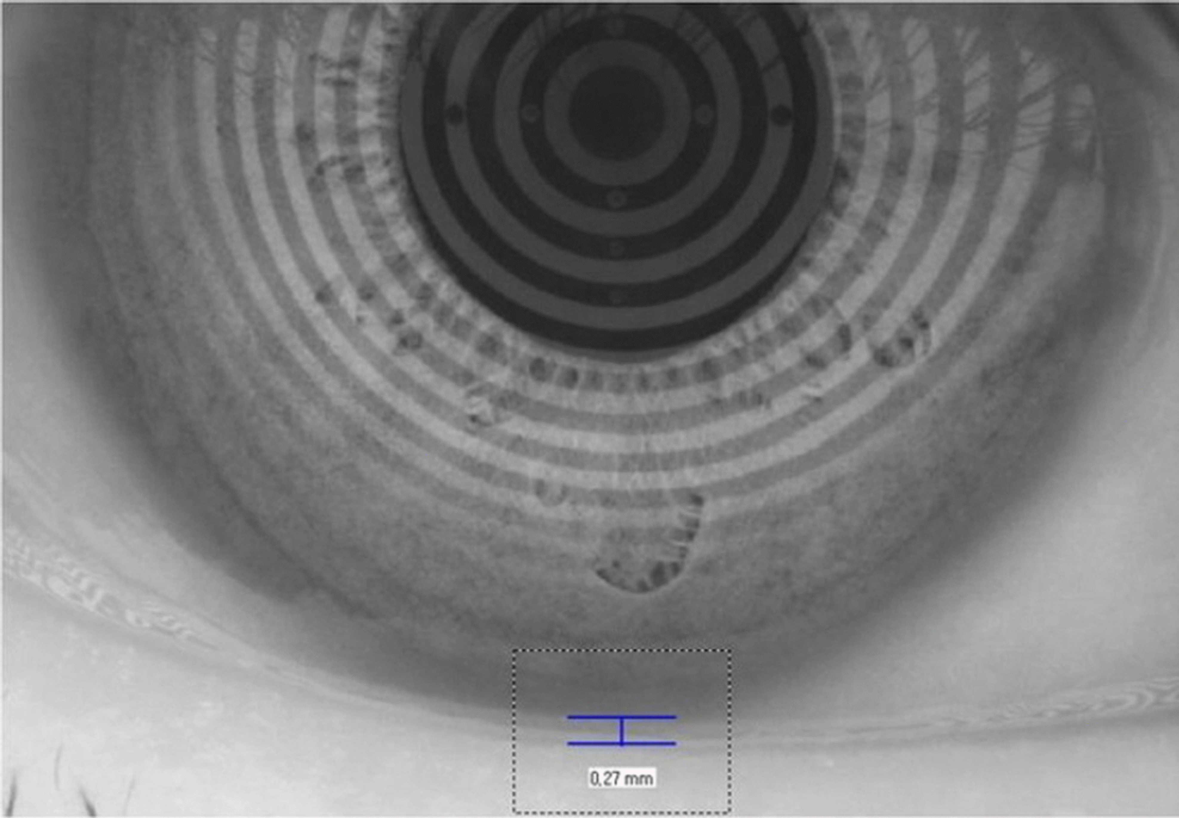
 | Figure 2.Measurement of tear meniscus height (TMH) using Keratograph 5M . The TMH was measured perpendicular to the lid margin at the pupil center. |
Table 1.
Demographic and clinical data of subjects enrolled in this study
Table 2.
Changes of Non-invasive Keratograph tear break-up time-first & average (NIKBUT-first & average), Non-invasive Keratograph tear meniscus height (NIK-TMH) over time in the patients
| Parameter of Keratograph | Preoperative day | Postoperative day | ||
|---|---|---|---|---|
| 1 day | 1 week | 1 month | ||
| NIKBUT-first (sec) | 10.74 ± 4.20 | 10.79 ± 4.91 | 6.39 ± 3.77 | 5.87 ± 2.79 |
| p-value* | 0.97 | <0.05 | <0.05 | |
| NIKBUT-average (sec) | 14.11 ± 3.60 | 14.59 ± 4.38 | 11.27 ± 5.21 | 11.28 ± 5.52 |
| p-value* | 0.70 | 0.36 | 0.57 | |
| NIK-TMH (mm) | 0.37 ± 0.15 | 0.35 ± 0.20 | 0.31 ± 0.16 | 0.30 ± 0.13 |
| p-value* | 0.58 | 0.16 | 0.09 | |


 PDF
PDF ePub
ePub Citation
 Print
Print


 XML Download