

 PDF
PDF ePub
ePub Citation
 Print
Print


Abstract
Purpose
To report the incidence of infraorbital nerve hypesthesia after inferior orbital wall fracture and reconstruction surgery and analyze the duration and factors to influence the occurence of the infraorbital nerve hypesthesia.
Methods
From March, 2001 to March, 2016, the medical records of 171 patients with isolated orbital floor fracture reconstructed with porous polyethylene or titanium mesh was analyzed retrospectively. Injury mechanism, fracture type, time interval to sur-gery, fracture size, type and thickness of implant were analyzed. Orbit computed tomography scan was performed at pre-operative and postoperative 6 weeks.
Results
Mean age was 30.4 years (male:female = 130:41). The mean time interval to surgery was 9.5 days. Incidence of in-fraorbital hypesthesia was 9.9% preoperatively, 38% in a week of surgery, 13.5% in 6 weeks and 5.8% in 6 months. Infraorbital hypesthesia lasts 20.5 weeks and the length of infraorbital canal was the only risk factor of persistent infraorbital hypesthesia.
Conclusions
Postoperative infraorbital nerve hypesthesia presents in a week in most patients. It last about 20.5 weeks, then mostly recovers in 6 months. This study will be useful to predict the clinical course of the patients with infraorbital nerve hypesthesia. Therefore, full explanation about the facial sense change is necessary for the patients with inferior orbital wall fracture.
Go to : 
References
1. Renzi G, Carboni A, Perugini M. . Posttraumatic trigeminal nerve impairment: a prospective analysis of recovery patterns in a series of 103 consecutive facial fractures. J Oral Maxillofac Surg. 2004; 62:1341–6.

2. Gierloff M, Seeck NG, Springer I. . Orbital floor re-construction with resorbable polydioxanone implants. J Craniofac Surg. 2012; 23:161–4.
3. Kruschewsky Lde S, Novais T, Daltro C. . Fractured orbital wall reconstruction with an auricular cartilage graft or absorbable polyacid copolymer. J Craniofac Surg. 2011; 22:1256–9.
4. Kontio R, Suuronen R, Salonen O. . Effectiveness of operative treatment of internal orbital wall fracture with polydioxanone implant. Int J Oral Maxillofac Surg. 2001; 30:278–85.
5. Folkestad L, Granström G. . A prospective study of orbital fracture sequelae after change of surgical routines. J Oral Maxillofac Surg. 2003; 61:1038–44.
6. Al-Sukhun J, Lindqvist C. . A comparative study of 2 implants used to repair inferior orbital wall bony defects: autogenous bone graft versus bioresorbable poly-L/DL-Lactide [P(L/DL)LA 70/30] plate. J Oral Maxillofac Surg. 2006; 64:1038–48.
7. Becker ST, Terheyden H, Fabel M. . Comparison of collagen membranes and polydioxanone for reconstruction of the orbital floor after fractures. J Craniofac Surg. 2010; 21:1066–8.
8. Beck-Broichsitter BE, Acar C, Kandzia C. . Reconstruction of the orbital floor with polydioxanone: a long-term clinical survey of up to 12 years. Br J Oral Maxillofac Surg. 2015; 53:736–40.
9. Brucoli M, Arcuri F, Cavenaghi R, Benech A. . Analysis of complications after surgical repair of orbital fractures. J Craniofac Surg. 2011; 22:1387–90.
10. Cai EZ, Koh YP, Hing EC. . Computer-assisted navigational surgery improves outcomes in orbital reconstructive surgery. J Craniofac Surg. 2012; 23:1567–73.
11. Jang KH, Kim NJ, Choung HK, Khwarg SI. . Orbital wall fracture repair: the results of early and delayed surgery. J Korean Ophthalmol Soc. 2016; 57:181–7.
12. Jeon C, Shin JH, Woo KI, Kim YD. . Porous polyethylene/titanium implants in the treatment of large orbital fractures. J Korean Ophthalmol Soc. 2009; 50:1133–40.
13. Yoon JS, Chung SA, Lee SY. . Repair of large posterior inferior wall fracture using medpo (R) channel sheet implant. J Korean Ophthalmol Soc. 2006; 47:1217–24.
14. Yang PJ, Chi NC, Choi GJ. . Comparison of sugical outcome be-tween early and delayed repair of orbital wall fracture. J Korean Ophthalmol Soc. 2003; 44:1278–84.
Go to :
 | Figure 1.Types of inferior orbital wall fractures in the images from the orbital CT scan. I: Fracture not involving the infraorbital ca-nal (I a: coronal view, I b: sagittal view). II: Fracture involving infraorbital canal and medial side from the canal. III: Fracture involving infraorbital canal and temporal side from the canal. IV: Large fracture involving infraorbital canal. V: Focal trap door fracture involving infraorbital canal. CT = computed tomography. |
 | Figure 2.Infraorbital canal length and angle meas-urement in the sagittal view of the contralateral or-bits from the orbital computed tomography (CT) scan. The length of the infraorbital canal and the angle between the inferior orbital wall and the in-fraorbital canal were measured in the sagittal CT image that the infraorbital canal was well vi-sualized on the assumption that the orbits are symmetric. |
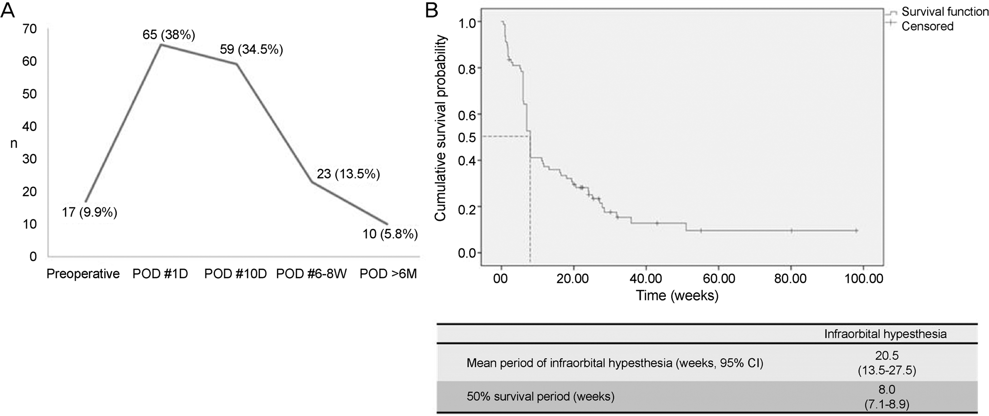 | Figure 3.Infraorbital nerve hypesthesia after inferior orbital wall fracture repair surgery. (A) Prevalence of infraorbital nerve hypesthesia. (B) Kaplan-Meier survival analysis for infraorbital hypesthesia following orbital fracture reconstruction surgery. POD = post operative days; CI = confidence interval; D =days; W = week(s); M = months. |
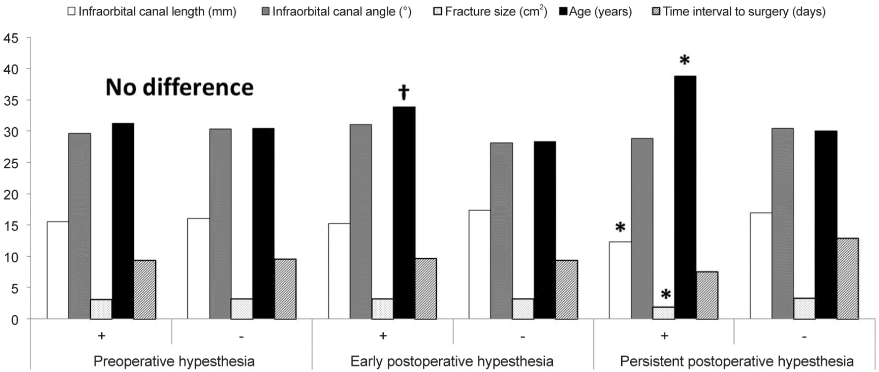 | Figure 4.Characteristics of the patients with infraorbital nerve hypesthesia. Early postoperative hypesthesia is a hypesthesia occurs in 10 days after reconstruction surgery. Persistent postoperative hypesthesia is a hypesthesia lasts more than 6 months. * p<0.05, Mann Whitney test; † p<0.05, Independent t-test. |
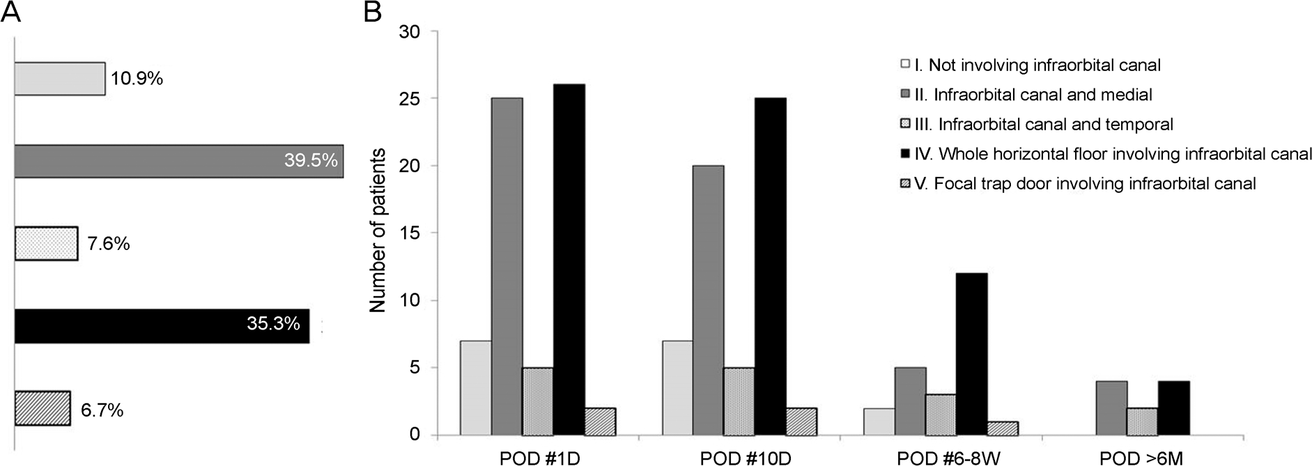 | Figure 5.Analysis according to the types of inferior orbital wall fracture. (A) Inferior orbital wall fracture types in the patients. (B) Patients with infraorbital nerve hypesthesia in postoperative status. POD = post operative days; D =days; W = week(s); M = months. |
Table 1.
Demographic characteristics of the patients with in-ferior orbital wall fracture
Table 2.
Factors to influence the occurrence of early and persistent infraorbital nerve hypesthesia in the patients with inferior orbital wall fracture
| Infraorbital nerve hypesthesia | Early | Persistent | ||||||
|---|---|---|---|---|---|---|---|---|
| B | S.E. | p-value | Exp (B) | B | S.E. | p-value | Exp (B) | |
| Infraorbital canal length (mm) | -0.427 | 0.217 | 0.048* | 0.652 | -0.423 | 0.195 | 0.030* | 0.655 |
| Infraorbital canal angle (°) | -0.153 | 0.161 | 0.342 | 0.858 | -0.304 | 0.175 | 0.082 | 0.738 |
| Time interval to surgery (days) | 0.338 | 0.426 | 0.427 | 1.402 | -0.092 | 0.145 | 0.526 | 0.912 |
| Fracture size > 2 cm2 | -1.031 | 1.463 | 0.481 | 0.357 | -7.843 | 7.575 | 0.300 | 0 |
| Age (years) | 0.116 | 0.220 | 0.597 | 1.123 | -0.122 | 0.119 | 0.306 | 0.885 |
| Preoperative infraorbital hypesthesia | - | -2.440 | 2.049 | 0.234 | 0.087 | |||
| Porous polyethylene with titanium mesh | 2.134 | 4.692 | 0.649 | 8.447 | ||||
| Sex | 1.378 | 5.420 | 0.799 | 3.968 | ||||
| Early hypesthesia | -0.189 | 1.788 | 0.916 | 0.828 | ||||
Table 3.
Overview of previous studies on perioperative hypesthesia of the patients with inferior orbital wall fracture
 XML Download