Abstract
Purpose
To compare the clinical results of short-term visual acuity and quality of vision after implantation of a yellow-tinted blue light-filtering intraocular lens (IOL) (Acrysof IQ® SN60WF) and an clear ultraviolet (UV) light filtering IOL (enVista TM MX60) in the same patient.
Methods
44 patients with bilateral cataract received an SN60WF in one eye and an MX60 in the other eye. All eyes were eval-uated for refraction power and uncorrected visual acuity (UCVA) at preoperative and 1, 3 months postoperatively. At post-operative 3 months, corrected visual acuity, quality of vision (OQASⅡ®), contrast sensitivity (CGT 2000®) and visual field (Humphrey Field Analyzer®), and subjective patients' response to the degree of brightness were evaluated. Furthermore, glisten-ing degree, intraocular stability, and posterior capsular opacification were examined.
Results
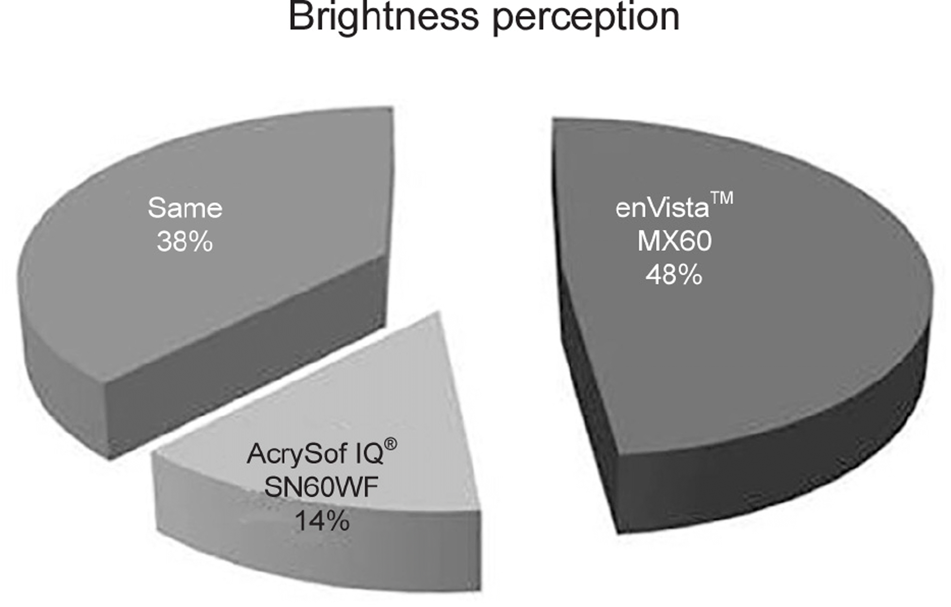
There were no significant differences in average refractive power or UCVA at 1 and 3 months ( p > 0.05) between the two groups. At 3 months after cataract surgery, the quality of vision according to OQASⅡ®, the contrast sensitivity according to CGT 2000® with the glare either on or off, and visual field; showed no difference between the two groups ( p > 0.05). Both IOLs had no glistening and posterior capsular opacity. The patients' response to the degree of brightness shows that MX60 (48.3%) has a higher degree of satisfaction.
Go to : 
References
1. Yoon KC, Mun GH, Kim SD, et al. Prevalence of eye diseases in South Korea: data from the Korea National Health and Nutrition Examination Survey 2008-2009. Korean J Ophthalmol. 2011; 25:421–33.

2. Putting BJ, van Best JA, Zweypfenning RC, et al. Spectral sensi-tivity of the blood-retinal barrier at the pigment epithelium for blue light in the 400-500 nm range. Graefes Arch Clin Exp Ophthalmol. 1961; 66:111–24.

3. Sparrow JR, Miller AS, Zhou J. Blue light-absorbing intraocular lens and retinal pigment epithelium protection in vitro. J Cataract Refract Surg. 1961; 66:111–24.

4. Ham WT Jr, Mueller HA, Sliney DH. Retinal sensitivity to damage from short wavelength light. Nature. 1961; 66:111–24.

5. Pollack A, Marcovich A, Bukelman A, Oliver M. Age-related mac-ular degeneration after extracapsular cataract extraction with intra-ocular lens implantation. Ophthalmology. 1961; 66:111–24.

6. Klein R, Klein BE, Wong TY, et al. The association of cataract and cataract surgery with the long-term incidence of age-related macul-opathy: the Beaver Dam eye study. Arch Ophthalmol. 2002; 120:1551–8.
7. Wang JJ, Klein R, Smith W, et al. Cataract surgery and the 5-year incidence of late-stage age-related maculopathy: pooled findings from the Beaver Dam and Blue Mountains eye studies. Ophthalmology. 1961; 66:111–24.
8. Mester U, Holz F, Kohnen T, et al. Intraindividual comparison of a blue-light filter on visual function: AF-1 (UY) versus AF-1 (UV) intraocular lens. J Cataract Refract Surg. 1961; 66:111–24.

9. Schmack I, Schimpf M, Stolzenberg A, et al. Visual quality assess-ment in patients with orange-tinted blue light-filtering and clear ul-traviolet light-filtering intraocular lenses. J Cataract Refract Surg. 1961; 66:111–24.

10. Zhu XF, Zou HD, Yu YF, et al. Comparison of blue light-filtering IOLs and UV light-filtering IOLs for cataract surgery: a meta-analysis. PLoS One. 2012; 7:e33013.

11. Werner L. Glistenings and surface light scattering in intraocular lenses. J Cataract Refract Surg. 1961; 66:111–24.

12. Colin J, Praud D, Touboul D, Schweitzer C. Incidence of glisten-ings with the latest generation of yellow-tinted hydrophobic acryl-ic intraocular lenses. J Cataract Refract Surg. 1961; 66:111–24.

13. Oshika T, Shiokawa Y, Amano S, Mitomo K. Influence of glisten-ings on the optical quality of acrylic foldable intraocular lens. Br J Ophthalmol. 1961; 66:111–24.

14. Colin J, Orignac I. Glistenings on intraocular lenses in healthy eyes: effects and associations. J Refract Surg. 1961; 66:111–24.

15. Dhaliwal DK, Mamalis N, Olson RJ, et al. Visual significance of glistenings seen in the AcrySof intraocular lens. J Cataract Refract Surg. 1961; 66:111–24.

16. Bae HW, Kim EK, Kim TI. Spherical aberration, contrast sensi-tivity and depth of focus with three aspherical intraocular lenses. J Korean Ophthalmol Soc. 1961; 66:111–24.

17. Kang IS, You IC, Park YG, Yoon KC. Comparison of visual func-tion among aspheric intraocular lenses. J Korean Ophthalmol Soc. 1961; 66:111–24.

18. Mun GH, Im SK, Park HY, Yoon KC. Comparison of visual func-tion between two aspheric intraocular lenses after microcoaxial cataract surgery. J Korean Ophthalmol Soc. 1961; 66:111–24.

19. Lee KH, Yoon MH, Seo KY, et al. Comparisons of clinical results after implantation of three aspheric intraocular lenses. J Korean Ophthalmol Soc. 1961; 66:111–24.

20. Kang MJ, Hwang HB, Chung SK. Effect of glistening-free intra-ocular lens on intraocular straylight. J Korean Ophthalmol Soc. 1961; 66:111–24.

21. Park YS, Ji YS, Yoon KC. Comparison of clinical long-term out-comes with two types of one-piece aspheric intraocular lenses after cataract surgery. J Korean Ophthalmol Soc. 1961; 66:111–24.

22. Colin J, Orignac I, Touboul D. Glistenings in a large series of hy-drophobic acrylic intraocular lenses. J Cataract Refract Surg. 1961; 66:111–24.

23. Chew EY, Sperduto RD, Milton RC, et al. Risk of advanced age-re-lated macular degeneration after cataract surgery in the Age-Related Eye Disease Study: AREDS report 25. Ophthalmology. 2009; 116:297–303.
24. Mainster MA, Turner PL. Blue-blocking IOLs decrease photo-reception without providing significant photoprotection. Surv Ophthalmol. 1961; 66:111–24.

25. Apple DJ, Mamalis N, Olson RJ, Kincaid MC. Intraocular lenses: evolution, designs, complications, and pathology. 1st ed.Baltimore: Williams & Wilkins;1989. p. 11–41.
26. Packer M, Rajan M, Ligabue E, Heiner P. Clinical properties of a novel, glistening-free, single-piece, hydrophobic acrylic IOL. Clin Ophthalmol. 1961; 66:111–24.

27. Packer M, Fry L, Lavery KT, et al. Safety and effectiveness of a glistening-free single-piece hydrophobic acrylic intraocular lens (enVista). Clin Ophthalmol. 1961; 66:111–24.

28. Heiner P, Ligabue E, Fan A, Lam D. Safety and effectiveness of a single-piece hydrophobic acrylic intraocular lens (enVista[R]) - re-sults of a European and Asian-Pacific study. Clin Ophthalmol. 1961; 66:111–24.
Go to : 
Table 1.
Patient demographics
|
Intraocular lens type |
p-value* | ||
|---|---|---|---|
| enVista TM MX60 | AcrySof IQ® SN60WF | ||
| Number of eyes | 44 | 44 | |
| OD:OS | 24:20 | 20:24 | |
| Sex (male:female) | 14:30 | 14:30 | |
| Mean age (years) | 70.0 ± 8.6 | 70.0 ± 8.6 | |
| Pre OP S.E. (D) | 0.55 ± 1.34 | 0.20 ± 1.94 | 0.370 |
| UCVA (Snellen) | 0.42 ± 0.17 | 0.46 ± 0.19 | 0.314 |
| Axial length (mm) | 23.23 ± 0.70 | 23.22 ± 0.66 | 0.954 |
| Intraocular lens power (D) | 21.30 ± 1.80 | 21.16 ± 1.70 | 0.716 |
| Foveal thickness measured by OCT (μ m) | 228.65 ± 24.73 | 229.29 ± 18.22 | 0.916 |
Table 2.
Refractive and visual outcomes at postoperative 1 month and 3 months
|
Intraocular lens type |
p-value* | |||
|---|---|---|---|---|
| enVista TM MX60 | AcrySof IQ® SN60WF | |||
| Postop 1 month | UCVA | 0.79 ± 0.17 | 0.77 ± 0.15 | 0.427 |
| S.E. (D) | 0.00 ± 0.49 | 0.09 ± 0.45 | 0.365 | |
| Postop 3 months | UCVA | 0.80 ± 0.16 | 0.78 ± 0.17 | 0.696 |
| BCVA | 0.96 ± 0.08 | 0.94 ± 0.09 | 0.434 | |
| S.E. (D) | -0.02 ± 0.53 | 0.07 ± 0.41 | 0.372 | |
Table 3.
Optical quality parameters measured by optical quality analysis system II® (OQAS II®) at postoperative 3 months
|
Intraocular lens type |
p-value* | ||
|---|---|---|---|
| enVista TM MX60 | AcrySof IQ® SN60WF | ||
| OSI | 1.28 ± 0.84 | 1.35 ± 1.04 | 0.727 |
| MTF cut-off value | 32.15 ± 9.08 | 31.98 ± 10.68 | 0.936 |
| Strehl ratio | 0.17 ± 0.05 | 0.16 ± 0.05 | 0.507 |
| VA100 | 1.08 ± 0.31 | 1.07 ± 0.36 | 0.322 |
| VA20 | 0.75 ± 0.25 | 0.74 ± 0.28 | 0.869 |
| VA9 | 0.44 ± 0.15 | 0.42 ± 0.15 | 0.539 |
Values are presented as mean ± standard deviation unless otherwise indicated. OSI = objective scatter index; MTF = modulation transfer function; VA100 = optical quality of the eye for 100% contrast conditions; VA20 = optical quality of the eye for 20% contrast conditions; VA9 = optical quality of the eye for 9% contrast conditions.
Table 4.
Visual field parameters measured by Humphrey field analyzer®: 24-2 SITA-Fast at postoperative 3 months
|
Intraocular lens type |
p-value* | ||
|---|---|---|---|
| enVista TM MX60 | AcrySof IQ® SN60WF | ||
| MD | -1.69 ± 0.89 | -1.67 ± 1.08 | 0.945 |
| PSD | 2.16 ± 0.86 | 2.41 ± 1.29 | 0.458 |
| Fovea threshold | 35.43 ± 1.96 | 35.09 ± 2.07 | 0.596 |
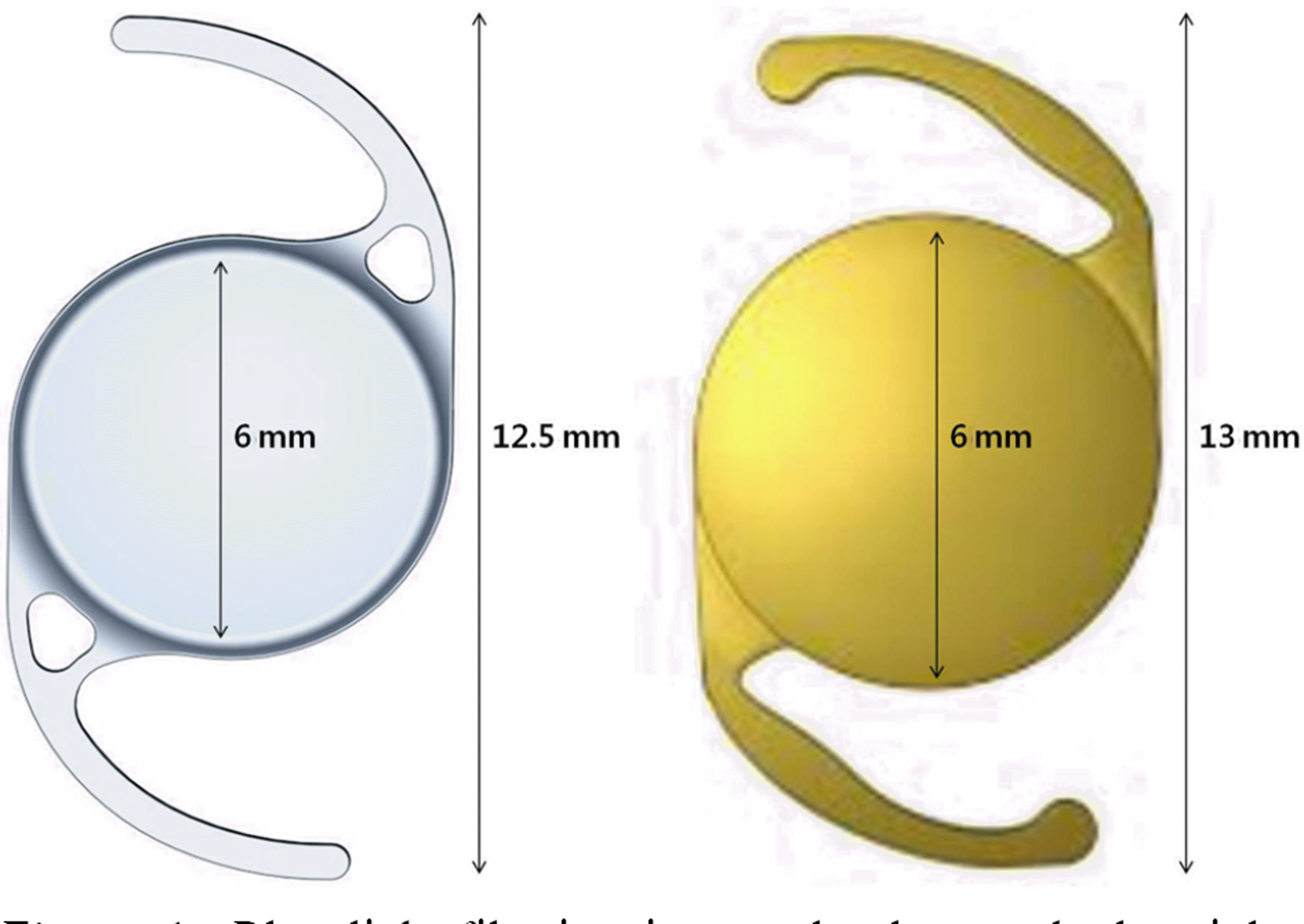
 | Figure 1.Blue light-filtering intraocular lens and ultraviolet (UV) light-filtering intraocular lens. The left lens is enVista TM MX60 and the right lens is AcrySof IQ® SN60WF. |
 | Figure 2.Contrast sensitivity test. The contrast sensitivity measurement using CGT-2000® was compared at (A) day, (B) twilight, and (C) night. Superior column tested when glare off, inferior column tested when glare on ( p < 0.05). |


 PDF
PDF ePub
ePub Citation
 Print
Print


 XML Download