

 PDF
PDF ePub
ePub Citation
 Print
Print


Abstract
Purpose
To determine whether the simultaneous recording of photopic electroretinography (ERG) and flash visual evoked potential (VEP) can predict the postoperative outcome in diabetic cases where massive vitreous hemorrhage precludes fundus observation.
Methods
The photopic ERG and flash VEP were recorded simultaneously on 20 eyes of 20 normal subjects, and 23 eyes of 23 patients who were diagnosed with Grade IV vitreous hemorrhage d/t diabetic retinopathy. Of the 23 patients, fellow eyes were diagnosed with proliferative diabetic retinopathy and they underwent pars plana vitrectomy after the test. Three groups were analyzed the responses of photopic ERG and flash VEP. Best corrected visual acuity was also checked before and after the surgery. After the 8 weeks after the vitrectomy, two groups were formed, based on the outcome of surgery and these two groups were analyzed the preoperative response of photopic ERG and flash VEP.
Results
When comparing the groups between proliferative diabetic retinopathy and normal eyes, Grade IV vitreous hemorrhage and fellow eyes, there was a statistically significant (p < 0.05) difference in a wave amplitude, a wave implicit time, b wave amplitude, b wave implicit time of photopic ERG and P2 peak time of flash VEP. In addition, a wave amplitude of photopic ERG showed the best predictive ability (area under receiver operating characteristic [AUROC] curve value of 0.88) when comparing improved visual acuity group to the unimproved visual acuity group.
Conclusions
Simultaneous recordings of photopic ERG and flash VEP showed the decreased function of retina and optic pathway on eyes with vitreous hemorrhage precluding inspection of the fundus. In addition, preoperative photopic ERG and flash VEP can safely predict the outcome of vitrectomy in dense vitreous hemorrhage of diabetics.
Go to : 
References
1. Goff MJ, McDonald HR, Johnson RN, et al. Causes and treatment of vitreous hemorrhage. Compr Ophthalmol Update. 2006; 7:97–111.
2. Ziemianski MC, McMeel JW, Franks EP. Natural history of vitreous hemorrhage in diabetic retinopathy. Ophthalmology. 1980; 87:306–12.

3. Francols J, Rouck AD. Electrodiagnosis, Toxic Agents and Vision: 15th I.S.C.E.V. Symposium Ghent, Belgium, June 20-23, 1977 (Documenta Ophthalmologica Proceedings Series) (2013-10–04). Netherlands: Springer. 1662; 193-201:203–9.
4. Freeman MH, Hirose T, Schepens CL. Vitreous Surgery and Advances in Fundus Diagnosis and Treatment. New York: Appleton-Century-Crofts;1977. p. 79–97.
5. Scherfig E, Tinning S, Edmund J, Trojaborg W. Visual evoked potential as prognostic factor for vitrectomy in diabetic eys. Acta Ophthalmol (Copenh). 1983; 61:778–87.
6. Kim HK, Kwon JY, Kim SH. Photopic electroretinogram in adult diabetics. J Korean Ophthalmol Soc. 1999; 40:121–7.
7. Youden WJ. Index for rating diagnostic tests. Cancer. 1950; 3:32–5.
8. Park SE, Sun HJ, Lee HJ, et al. The role of electroretinography in assessing the progression of diabetic retinopathy. J Korean Ophthalmol Soc. 2010; 51:693–9.
9. Bresnick GH, Palta M. Oscillatory potential amplitudes: Relation to severity of diabetic retinopathy. Arch Ophthalmol. 1987; 105:929–33.
10. Hiraiwa T, Horio N, Terasaki H, et al. Preoperative electroretinogram and postoperative visual outcome in patients with diabetic vitreous hemorrhage. Jpn J Ophthalmol. 2003; 47:307–11.
11. Yeom MI, Kim NE, Lee SJ, Park JM. Prognostic factors for neo-vascular glaucoma after vitrectomy in eyes with proliferative diabetic retinopathy. J Korean Ophthalmol Soc. 2015; 56:1229–35.
12. Scherfig E, Edmund J, Tinning S, Trojaborg W. Flash visual evoked potential as a prognostic factor for vitreous operations in diabetic eyes. Ophthalmology. 1984; 91:1475–9.
13. Skalka H, Holman J. Effect of pupillary dilatation in flash VER testing. Doc Ophthalmol. 1986; 63:321–4.
Go to :
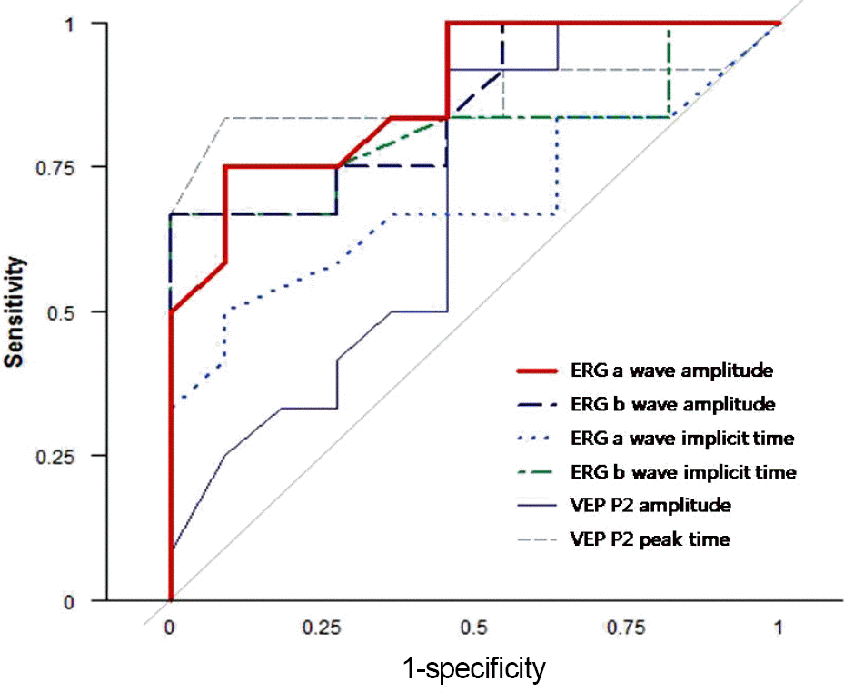 | Figure 1.Receiver operating characteristic curves of optical parameters for visual improvement. ERG = electroretinography; VEP = visual evoked potential. |
Table 1.
Characteristic per group
| Variable | Vit.Hm & PDR (N = 23) | Normal (N = 21) | Total (N = 44) | Comparison (p-value*) |
|---|---|---|---|---|
| Age (year) | 54.3 ± 8.92 | 53.67 ± 9.28 | 54.0 ± 8.99 | 0.817 |
| Sex (n, %) | ||||
| Male | 15 (65.22) | 12 (57.14) | 27 (61.36) | 0.811 |
| Female | 8 (34.78) | 9 (42.86) | 17 (38.64) |
Table 2.
Optical parameters per group
| Variable | Vit.Hm & PDR (N = 46) | Normal (N = 21) | Comparison (p-value*) |
|---|---|---|---|
| ERG | |||
| a amplitude (μV) | 11.78 ± 6.43 | 23.06 ± 3.30 | <0.001 |
| b amplitude (μV) | 17.70 ± 8.38 | 28.43 ± 3.70 | <0.001 |
| a implicit time (ms) | 19.05 ± 1.93 | 17.45 ± 3.33 | 0.006 |
| b implicit time (ms) | 48.24 ± 5.41 | 42.12 ± 1.29 | <0.001 |
| VEP | |||
| P2 amplitude (μV) | 13.09 ± 4.63 | 18.43 ± 8.96 | 0.024 |
| P2 peak time (ms) | 125.17 ± 12.26 | 120.88 ± 8.08 | 0.013 |
Table 3.
Optical parameters per group
| Variable | Vit.Hm (N = 23) | PDR (N = 23) | Comparison (p-value*) |
|---|---|---|---|
| ERG | |||
| a amplitude (μV) | 9.54 ± 4.91 | 14.01 ± 7.07 | 0.017 |
| b amplitude (μV) | 15.00 ± 6.71 | 20.39 ± 9.14 | 0.027 |
| a implicit time (ms) | 19.89 ± 1.95 | 18.20 ± 1.54 | 0.002 |
| b implicit time (ms) | 50.82 ± 5.71 | 45.66 ± 3.65 | 0.001 |
| VEP | |||
| P2 amplitude (μV) | 13.12 ± 2.63 | 13.07 ± 6.08 | 0.545 |
| P2 peak time (ms) | 129.65 ± 6.01 | 120.70 ± 15.15 | 0.029 |
Table 4.
Characteristic per improvement status in the Vitreous hemorrhage group
| Variable | Improved (N = 12) | Unimproved (N = 11) | Total (N = 23) | Comparison (p-value*) |
|---|---|---|---|---|
| Age (year) | 54.83 ± 9.31 | 53.73 ± 8.88 | 54.3 ± 8.92 | 0.774 |
| Sex | ||||
| Male | 6 (50) | 9 (81.82) | 15 (65.22) | 0.245 |
| Female | 6 (50) | 2 (18.18) | 8 (34.78) | |
| DM duration (year) | 14.33 ± 6.65 | 16 ± 7.64 | 15.13 ± 7.03 | 0.582 |
| Cataract | ||||
| O | 6 (50) | 8 (72.73) | 14 (60.87) | 0.491 |
| X | 6 (50) | 3 (27.27) | 9 (39.13) | |
| Photocoagulation | ||||
| O | 6 (50) | 6 (54.55) | 12 (52.17) | 1 |
| X | 6 (50) | 5 (45.45) | 11 (47.83) |
Table 5.
Optical parameters per group
| Variable | Improved (N = 12) | Unimproved (N = 11) | Comparison (p-value*) |
|---|---|---|---|
| ERG | |||
| a amplitude (μV) | 12.35 ± 4.94 | 6.48 ± 2.54 | 0.002 |
| b amplitude (μV) | 19.06 ± 6.71 | 10.58 ± 2.88 | 0.001 |
| a implicit time (ms) | 20.58 ± 2.19 | 19.14 ± 1.36 | 0.122 |
| b implicit time (ms) | 53.9 ± 5.22 | 47.46 ± 4.23 | 0.011 |
| VEP | |||
| P2 amplitude (μV) | 14.17 ± 1.39 | 11.97 ± 3.21 | 0.123 |
| P2 peak time (ms) | 126.21 ± 6.35 | 133.41 ± 2.31 | 0.003 |
Table 6.
Retinal finding as vitrectomy
| Variable Normal macular | Improved (N = 12) 6 | Unimproved (N = 11) 0 |
|---|---|---|
| Diabetic maculopathy | 4 | 4 |
| Fibrovascular membranes | 1 | 3 |
| Macular traction/detachment | 1 | 4 |
Table 7.
Predictive performance of optical parameter for visual improvement
 XML Download