Abstract
Purpose
To report on a patient with a brown pigmented mass in the anterior chamber suspected of a granuloma caused by a metallic foreign body and to review the relevant literature.
Case summary
A 63-year-old man presented with blurring in his right eye, which had initially began several years prior. Concentric corneoscleral brown pigmentation about 2 mm in diameter was found in the superonasal limbal area. A rectangular parallelepiped mass was observed in the superonasal anterior chamber, which was an even brown color with a small white por-tion, smooth-surfaced, and non-vascularized. The pupil was oval and dragged superonasally, possibly due to mild compression of the iris caused by the mass. In pre-enhanced orbital computed tomography, a round high signal intensity with a diameter of 3.0 mm was found in the superonasal anterior segment. Though excisional biopsy using the lamellar scleral flap was considered for exact diagnosis, this was not performed considering the clinical features are more indicative of granuloma than iris melanoma. Neither changes in the size of the mass nor the shape of the pupil were observed during the follow up period until 19 months after the first visit.
Conclusions
When a pigmented mass in the anterior chamber is detected, benign and malignant iris tumors and granulomas should be considered for a differential diagnosis. The patient’s exact past medical history and clinical features differentiating ma-lignant and benign masses are important for proper diagnosis due to the difficulty in obtaining tissue diagnoses in some cases. J Korean Ophthalmol Soc 2017;58(2):230-234
References
1. Shields CL, Kancherla S, Patel J. . Clinical survey of 3680 iris tumors based on patient age at presentation. Ophthalmology. 2012; 119:407–14.

2. Sala-Puigdollers A, Rodrígúez-de la Rúa E, Saornil MA. . Combined choroidal biopsy and cytology for diagnosis of intra-ocular tumour. Arch Soc Esp Oftalmol. 2013; 88:365–8.

3. Shields CL, Shields PW, Manalac J. . Review of cystic and sol-id tumors of the iris. Oman J ophthalmol. 2013; 6:159–64.

4. Toth J. Clinical signs and differential diagnosis of iris melanoma. Magy Onkol. 2004; 49(153-5):158–9.
5. Goldsmith LA, Askin FB, Chang AE. . Diagnosis and treat-ment of early melanoma: NIH consensus development panel on early melanoma. JAMA. 1992; 268:1314–9.
6. Jain V, Dabir S, Shome D. . Aspergillus iris granuloma: a case report with review of literature. Surv Ophthalmol. 2009; 54:286–91.

7. Womack LW, Liesegang TJ. Complications of herpes zoster ophthalmicus. Arch Ophthalmol. 1983; 101:42–5.

8. Allen JH. The pathology of ocular leprosy. II. Miliary lepromas of the iris. Am J Ophthalmol. 1966; 61((5 Pt 2)):987–92.
9. Ocampo VV Jr, Foster CS, Baltatzis S.Surgical excision of iris nodules in the management of sarcoid uveitis. Ophthalmology. 2001; 108:1296–9.

10. Pinto A, Brunese L, Daniele S. . Role of computed tomography in the assessment of intraorbital foreign bodies. Semin Ultrasound CT MR. 2012; 33:392–5.

11. Albert DM, Miller JW, Azar DT, Blodi B. Albert & Jakobiec's Principles & Practice of Ophthalmology. 3rd ed. Phildadelphia: Elsevier Science Health Science div;2007. p. 5143.
Figure 1.
Slit lamp examination of the patient. (A) Slit-lamp examination revealed oval shaped pupil (arrow) drawn to superonasally and brown pigmentation (arrowhead) on the superonasal peripheral cornea, which is concentric and centered on a limbal point. (B, C) The superonasal pigmentation consists of two layers, of which the superficial layer was located in the anterior stromal level with diameter of 1 mm (arrows) and the deep layer was in the posterior stromal level with diameter of 2 mm (arrowheads). (D) Gonioscopy visualized rectangular parallelepiped mass (2 mm wide × 0.6 mm height × 0.3 mm depth, arrow) in the superonasal anterior chamber. The mass consists of a mainly even-brown-colored double-hump-shaped portion and a small white portion be-tween the two humps. The surface of the mass was smooth and non-vascularized.

Figure 2.
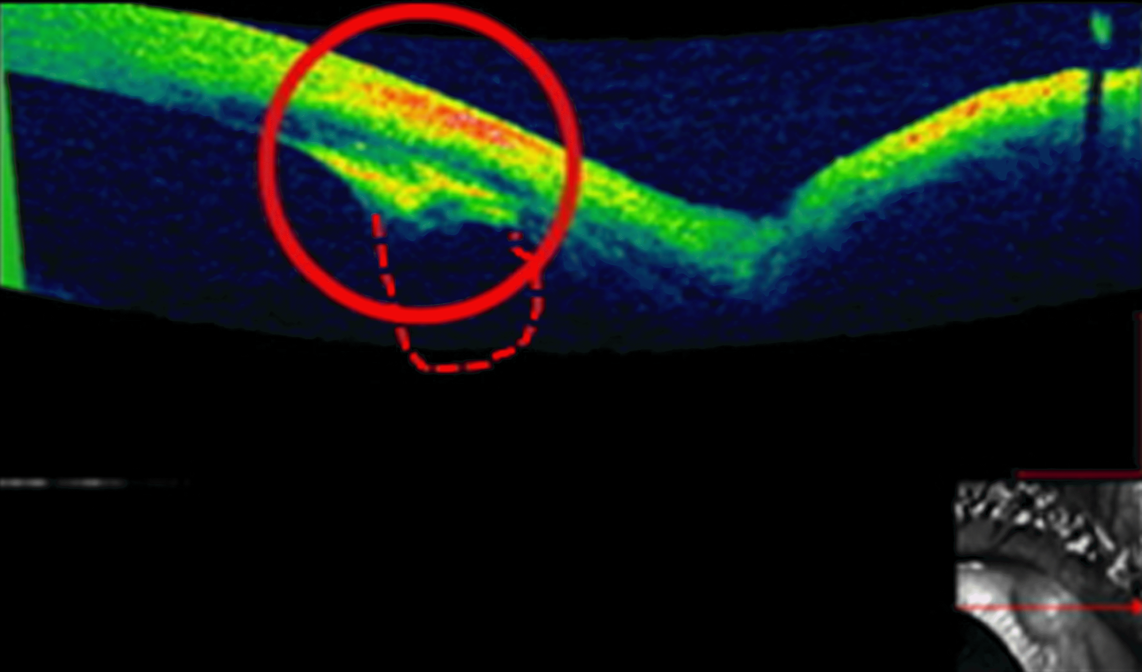
Anterior segment optical coherence tomography of the patient. The red circle showed two-layered separate in-creased density in the anterior stroma and posterior stroma in-cluding endothelium. A mass with increased density originates from the posterior stroma and endothelium of the cornea. However, the mass area inside the anterior chamber, which was observed by gonioscopy, was not visualized by anterior segment optical coherence tomography possibly due to the rapid signal attenuation of the light by the mass. The dotted line simulates imaginary outline of the mass considering the shape of the mass seen by gonioscopy.


 PDF
PDF ePub
ePub Citation
 Print
Print


 XML Download