Abstract
Purpose
To report the treatment results of a frontotemporal dermoid cyst with a cutaneous fistula and sinus tract that caused re-current periorbital cellulitis in a child.
Case summary
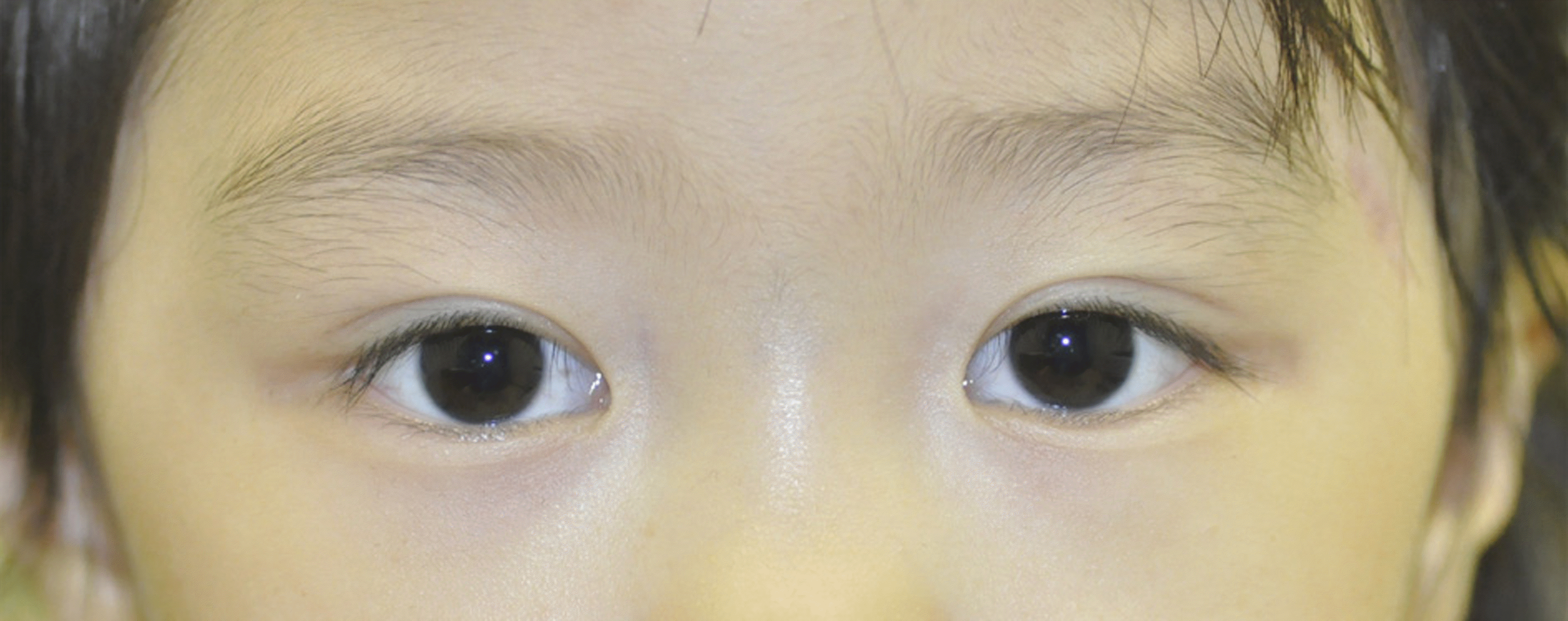
A 4-year-old girl who presented with left orbital swelling and tenderness visited our hospital. She had a cuta-neous fistula with a small amount of purulent discharge at the left frontotemporal area. Orbital computed tomography scans showed a well-defined low density lesion in the fronto-zygomatic suture, and there was a bony defect in the left greater wing of the sphenoid bone of the orbit. Orbital magnetic resonance imaging showed a cutaneous fistula and sinus tract that extended in-to the middle cranial fossa. The patient was treated with intravenous antibiotics until the inflammation was resolved. Surgery was performed to remove the dermoid cyst with sinus tract. After surgery, there was no evidence of recurrence, and complications in-cluded neurologic and ophthalmic symptoms.
Go to : 
References
1. Sherman RP, Rootman J, Lapointe JS. Orbital dermoids: clinical presentation and management. Br J Ophthalmol. 1984; 68:642–52.

3. Pollard ZF, Calhoun J. Deep orbital dermoid with draining sinus. Am J Ophthalmol. 1975; 79:310–3.

4. Bartlett SP, Lin KY, Grossman R, Katowitz J. The surgical man-agement of orbitofacial dermoids in the pediatric patient. Plast Reconstr Surg. 1993; 91:1208–15.

5. Bonavolontà G, Tranfa F, de Conciliis C, Strianese D.Dermoid cyst: 16 year survey. Ophthal Plast Reconstr Surg. 1995; 11:187–92.
6. Borley WE. Dermoid (OIL) cyst of the orbit: Report of a case. Am J Ophthalmol. 1939; 22:1355–9.
7. Pensler JM, Bauer BS, Naidich TP. Craniofacialdermoids. Plast Reconstr Surg. 1988; 82:953–8.
8. Hong SW. Deep frontotemporal dermoid cyst presenting as a dis-charging sinus: a case report and review of literature. Br J Plast Surg. 1998; 51:255–7.

9. Hönig JF. A de novo discharging sinus of the fronto-orbital suture: a rare presentation of a dermoid cyst. J Craniofac Surg. 1998; 9:536–8.
10. Posnikc JC, Bortoluzzi P, Armstrong DC, Drake JM. Intracranial nasal dermoid sinus cyst: computed tomographic scan findings and surgical results. Plast Reconstr Surg. 1994; 93:745–54.
Go to : 
 | Figure 1.Clinical photographs of the patient. (A) Cutaneous fistula with purulent discharge at left frontotemporal area. (B) Lateral view. |
 | Figure 2.Computed tomography (CT) scan of patient. (A) Axial CT scan (bone window) show bony defect in the left greater wing of sphenoid which is interconnected with the temporal fossa. (B) Coronal CT scan showing well defined low density lesion in fron-to-zygomatic suture. |
 | Figure 3.Magnetic resonance (MR) images of the patient. (A) Axial T2-weighted MR image showing cutaneous fistula and sinus tract extended into the middle cranial fossa in the left lateral orbital area. (B) Coronal T2-weighted MR image showing sinus tract that is well defined and in high signal lesion. |
 | Figure 4.The photographs of histopathological findings. (A) A dermal cyst which is lined by squamous epithelium is seen (hematoxylin-eosin stain, original magnification ×20). (B) The wall of the cyst has associated sebaceous glands and hair follicles (hematoxylin-eosin stain, original magnification ×20). |


 PDF
PDF ePub
ePub Citation
 Print
Print


 XML Download