

 PDF
PDF ePub
ePub Citation
 Print
Print


Abstract
Purpose
To analyze the effect of stereopsis and contrast sensitivity on the quality of life and to evaluate the relationship be-tween integrated binocular visual field (IVF) and binocular visual function in bilateral normal tension glaucoma (NTG) patients.
Methods
Stereopsis and contrast sensitivity tests were performed and compared among 44 NTG patients and 32 normal subjects. The IVF was integrated using the best location method. The correlation between visual function and subscales of the National Eye Institute Visual Function Questionnaire 25 (NEI VFQ-25) was evaluated using univariate linear regression.
Results
Stereopsis and contrast sensitivity for the bilateral NTG patients were decreased compared to the normal controls. Stereopsis and contrast sensitivity exhibited a significant correlation with social functions related to vision and color vision among subscales of NEI VFQ-25. IVF mean deviation (MD) and better eye MD showed a significant correlation with stereopsis and contrast sensitivity, while worse eye MD showed no association.
Go to : 
References
1. Gutierrez P, Wilson MR, Johnson C. . Influence of glaucoma-tous visual field loss on health-related quality of life. Arch Ophthalmol. 1997; 115:777–84.

2. Janz NK, Wren PA, Lichter PR. . Quality of life in newly diag-nosed glaucoma patients: The Collaborative Initial Glaucoma Treatment Study. Ophthalmology. 2001; 108:887–97.
3. McKean-Cowdin R, Wang Y, Wu J. . Impact of visual field loss on health-related quality of life in glaucoma: the Los Angeles Latino Eye Study. Ophthalmology. 2008; 115:941–8.e1.
4. Parrish RK 2nd, Gedde SJ, Scott IU. . Visual function and qual-ity of life among patients with glaucoma. Arch Ophthalmol. 1997; 115:1447–55.
5. Friedman DS, Freeman E, Munoz B. . Glaucoma and mobility performance: the Salisbury Eye Evaluation Project. Ophthalmology. 2007; 114:2232–7.
6. Gupta N, Krishnadev N, Hamstra SJ, Yücel YH. Depth perception deficits in glaucoma suspects. Br J Ophthalmol. 2006; 90:979–81.
7. Ramulu PY, West SK, Munoz B. . Glaucoma and reading speed: the Salisbury Eye Evaluation project. Arch Ophthalmol. 2009; 127:82–7.
8. McKendrick AM, Sampson GP, Walland MJ, Badcock DR. Contrast sensitivity changes due to glaucoma and normal aging: low-spatial-frequency losses in both magnocellular and parvocel-lular pathways. Invest Ophthalmol Vis Sci. 2007; 48:2115–22.
9. Sung MS, Park SW. Spatial contrast sensitivity for the diagnosis of glaucoma. Journal of The Korean Glaucoma Society. 2015; 4:14–20.
10. Chang JH, Chun BY, Shin JP. The stereopic acuity in patients with unilateral or bilateral visual field defects. J Korean Ophthalmol Soc. 2014; 55:734–9.
11. Junemann AG, Horn FK, Martus P, Korth M. The full-field tempo-ral contrast sensitivity test for glaucoma: influence of cataract. Graefes Arch Clin Exp Ophthalmol. 2000; 238:427–32.
12. Klein J, Pierscionek BK, Lauritzen J. . The effect of cataract on early stage glaucoma detection using spatial and temporal contrast sensitivity tests. PLoS One. 2015; 10:e0128681.
13. Kim CS, Seong GJ, Lee NH. . Prevalence of primary open-angle glaucoma in central South Korea the Namil study. Ophthalmology. 2011; 118:1024–30.
14. Kang BW, Ji YS, Park SW. Analysis of factors related of location of initial visual field defect in normal tenstion glaucoma. J Korean Ophthalmol Soc. 2011; 52:1478–84.
15. Heo JW, Yoon HS, Shin JP. . A validation and reliability study of the Korean version of National Eye Institute Visual Function Questionnaire 25. J Korean Ophthalmol Soc. 2010; 51:1354–67.
16. Crabb DP, Viswanathan AC, McNaught AI. . Simulating bin-ocular visual field status in glaucoma. Br J Ophthalmol. 1998; 82:1236–41.
17. Nelson-Quigg JM, Cello K, Johnson CA.Predicting binocular vis-ual field sensitivity from monocular visual field results. Invest Ophthalmol Vis Sci. 2000; 41:2212–21.
18. Chun YS, Park IK. Comparision of mean deviation between in-tegrated binocular visual field and monocular visual field. J Korean Ophthalmol Soc. 2013; 54:919–26.
19. Lee DI, Park IK, Jeong JH, Chun YS. Quality of life according to location of integrated binocular visual field defect in nor-mal-tension-glaucoma patients. J Korean Ophthalmol Soc. 2016; 57:86–97.
20. Anderson DR. Kist K, editor. The single field printout with Statpac analysis. Automated Static Perimetry. 2nd ed. St. Louis: Mosby;1992. p. chap. 5.
21. Bergua A, Horn FK, Martus P. . Stereoscopic visual evoked po-tentials in normal subjects and patients with open-angle glaucomas. Graefes Arch Clin Exp Ophthalmol. 2004; 242:197–203.
22. Parisi V. Impaired visual function in glaucoma. Clin Neurophysiol. 2001; 112:351–8.
23. Derefeldt G, Lennerstrand G, Lundh B. Age variations in normal human contrast sensitivity. Acta Ophthalmol (Copenh). 1979; 57:679–90.
24. Livingstone MS, Hubel DH. Psychophysical evidence for separate channels for the perception of form, color, movement, and depth. J Neurosci. 1987; 7:3416–68.
25. Maunsell JH, Van Essen DC. Functional properties of neurons in middle temporal visual area of the macaque monkey. I. Selectivity for stimulus direction, speed, and orientation. J Neurophysiol. 1983; 49:1127–47.
26. Schiller PH. The central visual system. Vision Res. 1986; 26:1351–86.
27. Klistorner AI, Graham SL. Early magnocellular loss in glaucoma demonstrated using the pseudorandomly stimulated flash visual evoked potential. J Glaucoma. 1999; 8:140–8.
28. . . Loss of neurons in magnocel-lular and parvocellular layers of the lateral geniculate nucleus in glaucoma. Arch Ophthalmol. 2000; 118:378–84.
29. Yücel YH, Zhang Q, Weinreb RN. . Effects of retinal ganglion cell loss on magno-, parvo-, koniocellular pathways in the lateral geniculate nucleus and visual cortex in glaucoma. Prog Retin Eye Res. 2003; 22:465–81.
30. Derrington AM, Lennie P. Spatial and temporal contrast sensitiv-ities of neurones in lateral geniculate nucleus of macaque. J Physiol. 1984; 357:219–40.
Go to :
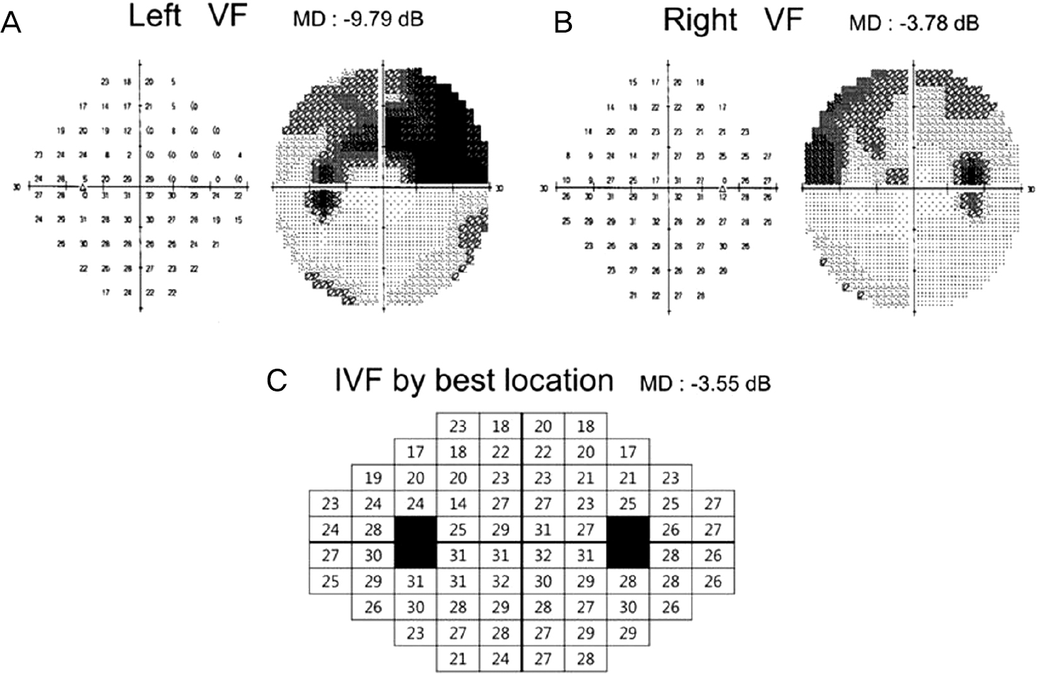 | Figure 1.Integrated visual field (IVF) of normal tension glaucoma. Schematic representation of the left (A), right (B) and IVF by best location (C). VF = visual field; MD = mean deviation; dB = decibels. |
Table 1.
Demographic data for normal tension glaucoma (NTG) and control
| NTG (n = 44) | Control (n = 32) | p-value* | |
|---|---|---|---|
| Mean age (years) | 59.02 ± 14.47 | 57.56 ± 14.07 | 0.672 |
| Gender (male/female) | 25/19 | 22/10 | 0.297 |
| Visual acuity (logMAR) | |||
| Better eye | 0.11 ± 0.11 | 0.09 ± 0.09 | 0.568 |
| Worse eye | 0.15 ± 0.11 | 0.13 ± 0.08 | 0.607 |
| Intraocular pressure (mmHg) | |||
| Higher eye | 14.02 ± 2.66 | 13.78 ± 2.35 | 0.683 |
| Lower eye | 12.93 ± 2.64 | 12.62 ± 2.56 | 0.614 |
Table 2.
Clinical data for normal tension glaucoma (NTG) and control
| NTG (n = 44) | Control (n = 32) | p-value* | |
|---|---|---|---|
| MD of visual field (dB) | |||
| Binocular (IVF) | -3.99 ± 6.29 | 2.29 ± 1.5 | <0.001 |
| Better eye | -5.04 ± 6.71 | 0.97 ± 3.5 | <0.001 |
| Worse eye | -9.52 ± 8.39 | -0.02 ± 2.4 | <0.001 |
| Stereopsis (sec) | 174.09 ± 185.18 | 44.77 ± 11.71 | <0.001 |
| Contrast sensitivity test (Photopic, log) | |||
| 3 cpd | 1.67 ± 0.18 | 1.76 ± 0.17 | 0.039 |
| 6 cpd | 1.90 ± 0.20 | 2.03 ± 0.18 | 0.006 |
| 12 cpd | 1.57 ± 0.29 | 1.82 ± 0.18 | <0.001 |
| 18 cpd | 1.15 ± 0.25 | 1.42 ± 0.15 | <0.001 |
| Contrast sensitivity test (Mesopic, log) | |||
| 3 cpd | 1.67 ± 0.20 | 1.74 ± 0.14 | 0.129 |
| 6 cpd | 1.85 ± 0.20 | 2.01 ± 0.14 | <0.001 |
| 12 cpd | 1.49 ± 0.31 | 1.69 ± 0.18 | 0.002 |
| 18 cpd | 1.04 ±0.27 | 1.29 ± 0.18 | <0.001 |
Table 3.
Correlations of National Eye Institute Visual Function Questionnaire (NEI-VFQ-25) subscale score with clinical visual function tests
| NEI-VFQ-25 subscale | Score | Stereopsis |
Photopic constrast sensitivity test |
Mesopic constrast sensitivity test |
||||||
|---|---|---|---|---|---|---|---|---|---|---|
| 3 cpd | 6 cpd | 12 cpd | 18 cpd | 3 cpd | 6 cpd | 12 cpd | 18 cpd | |||
| General health | 36.58 | -0.25 | 0.21 | 0.12 | 0.28 | 0.02 | 0.36 ( p = 0.023*) | 0.01 | 0.35 ( p = 0.026*) | 0.04 |
| General VA | 66.50 | -0.33( p = 0.039*) | 0.08 | 0.03 | 0.08 | -0.01 | 0.09 | 0.02 | -0.11 | -0.09 |
| Ocular pain | 80.79 | -0.11 | 0.05 | -0.16 | -0.01 | -0.04 | 0.14 | -0.05 | -0.03 | 0.15 |
| Near acuity | 81.70 | -0.23 | 0.10 | 0.01 | 0.08 | 0.02 | 0.11 | 0.01 | -0.10 | 0.07 |
| Distance acuity | 83.94 | -0.35( p = 0.027*) | 0.14 | 0.15 | 0.16 | 0.15 | 0.27 | 0.04 | 0.02 | 0.15 |
| Social functioning | 91.46 | -0.37 ( p = 0.016*) | 0.21 | 0.28 | 0.34 ( p = 0.034*) | 0.32 ( p = 0.047*) | 0.25 | 0.24 | 0.22 | 0.40 ( p = 0.010*) |
| Mental health | 77.59 | -0.18 | 0.12 | 0.05 | 0.08 | -0.15 | 0.19 | 0.04 | 0.01 | 0.04 |
| Role difficulties | 74.35 | -0.29 | 0.08 | 0.02 | 0.11 | 0.01 | 0.31 | 0.15 | 0.05 | 0.08 |
| Dependency | 88.88 | -0.12 | -0.03 | 0.16 | 0.10 | -0.09 | 0.17 | 0.13 | -0.03 | -0.01 |
| Driving | 82.01 | -0.09 | -0.29 | 0.04 | 0.01 | 0.01 | -0.14 | 0.10 | 0.23 | 0.28 |
| Color vision | 93.90 | -0.45 ( p = 0.003*) | 0.27 | 0.28 | 0.34 ( p = 0.033*) | 0.20 | 0.25 | 0.25 | 0.26 | 0.36 ( p = 0.022*) |
| Peripheral vision | 90.24 | -0.21 | 0.34( p = 0.032* | 0.22) | 0.24 | 0.20 | 0.25 | 0.26 | 0.10 | 0.30 |
| Composite score | 82.94 | -0.40 ( p= 0.009*) | 0.23 | 0.12 | 0.20 | 0.11 | 0.25 | 0.12 | 0.02 | 0.20 |
Table 4.
Correlation coefficient of a univariate linear regression between visual function test and MDs
 XML Download