

 PDF
PDF ePub
ePub Citation
 Print
Print


Abstract
Purpose
To report three cases of glial hyper-proliferation after autologous internal limiting membrane (ILM) transplantation in idiopathic large macular holes.
Case Summary
Three eyes with full thickness macular holes >500 µm underwent autologous ILM transplantation. After surgery, the macular hole was closed and foveal contour was U-shaped. Optical coherence tomography revealed long-lasting proliferation of glial cells in the fovea after the hole closure. This glial proliferation continued for 6 months, with improved visual acuity, and bump-like features of the fovea.
Figures and Tables
Figure 1
A Case of 67-year-old woman with full thickness macular hole. Two weeks after autologous internal limiting membrane transplantation, optical coherence tomography images showed a glial tissues in the fovea without a complete hole closure. The long-lasting proliferation of glial cells (yellow arrows) resulted in the closure of hole, but the abnormal contour of fovea was postoperatively observed from 3 months to 24 months. The defects of ellipsoid zone (black arrowheads) and external limiting membrane (white arrowheads) remained. The foveal depigmentation was seen in the postoperative fundus photographs.
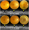
Figure 2
A case of 70-year-old woman with full thickness macular hole. One month after autologous internal limiting membrane transplantation, the closure of the hole and glial tissues in fovea (yellow arrow) were observed in optical coherence tomography. Foveal depigmentation was seen in fundus photography. At 6 months, the protrusion of fovea developed. The defects of ellipsoid zone (black arrowheads) and external limiting membrane (white arrowheads) remained.
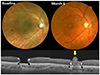
Figure 3
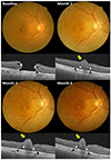
A case of 75-year-old woman with full thickness macular hole. One month after autologous internal limiting membrane transplantation, irregular contour of fovea (yellow arrows) was detected in optical coherence tomography. At 3 months, the bump-like fovea developed. The depigmentation of fovea was seen in postoperative fundus photograph. The defects of ellipsoid zone (black arrowheads) and external limiting membrane (white arrowheads) remained. And it lasted until 6 months.
References
1. Johnson RN, Gass JD. Idiopathic macular holes. Observations, stages of formation, and implications for surgical intervention. Ophthalmology. 1988; 95:917–924.
2. la Cour M, Friis J. Macular holes: classification, epidemiology, natural history and treatment. Acta Ophthalmol Scand. 2002; 80:579–587.
3. Brooks HL Jr. Macular hole surgery with and without internal limiting membrane peeling. Ophthalmology. 2000; 107:1939–1948. discussion 1948-9.
4. Salter AB, Folgar FA, Weissbrot J, Wald KJ. Macular hole surgery prognostic success rates based on macular hole size. Ophthalmic Surg Lasers Imaging. 2012; 43:184–189.
5. Michalewska Z, Michalewski J, Adelman RA, Nawrocki J. Inverted internal limiting membrane flap technique for large macular holes. Ophthalmology. 2010; 117:2018–2025.
6. Kim KH, Jung JW, Park SW, et al. Autologous transplantation of internal limiting membrane for the treatment of large macular hole. J Korean Ophthalmol Soc. 2015; 56:1899–1905.
7. Wender J, Iida T, Del Priore LV. Morphologic analysis of stage 3 and stage 4 macular holes: implications for treatment. Am J Ophthalmol. 2005; 139:1–10.
8. Stec LA, Ross RD, Williams GA, et al. Vitrectomy for chronic macular holes. Retina. 2004; 24:341–347.
9. De Novelli FJ, Preti RC, Ribeiro Monteiro ML, et al. Autologous internal limiting membrane fragment transplantation for large, chronic, and refractory macular holes. Ophthalmic Res. 2015; 55:45–52.
10. Smiddy WE, Feuer W, Cordahi G. Internal limiting membrane peeling in macular hole surgery. Ophthalmology. 2001; 108:1471–1476. discussion 1477-8.
11. Costa RA, Cardillo JA, Morales PH, et al. Optical coherence tomography evaluation of idiopathic macular hole treatment by gas-assisted posterior vitreous detachment. Am J Ophthalmol. 2001; 132:264–266.
12. Oh J, Yang SM, Choi YM, et al. Glial proliferation after vitrectomy for a macular hole: a spectral domain optical coherence tomography study. Graefes Arch Clin Exp Ophthalmol. 2013; 251:477–484.
13. Imai M, Iijima H, Gotoh T, Tsukahara S. Optical coherence tomography of successfully repaired idiopathic macular holes. Am J Ophthalmol. 1999; 128:621–627.
 XML Download