

 PDF
PDF ePub
ePub Citation
 Print
Print


Abstract
Purpose
To investigate the correlation between 24-hour ambulatory blood pressure (BP) monitoring and peripapillary retinal vessel width and visual field (VF) defect progression in normal tension glaucoma (NTG) patients.
Methods
All patients were classified by 24-hour ambulatory BP monitoring as non-dipper (nocturnal dip < 10%) and dipper (nocturnal dip ≥ 10%) group. Vessel diameter, mean deviation (MD) value by VF test and VF progression from Glaucoma Progression Analysis (GPA) were compared among non-dipper and dipper groups.
Results
Retinal arterial diameter was wider in the non-dipper group compared to the dipper group (p = 0.015), while retinal venous diameter had no significant relationship between the two groups (p = 0.131). The MD value at baseline and 2 years after was worse in the non-dipper group than the dipper group, respectively (p = 0.006, p = 0.030). But, there was no significant relationship between nocturnal dip and GPA progression (p = 0.658).
Figures and Tables
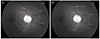 | Figure 1The geometric measurement of the retinal vessels. The 1 disc-diameter is marked as the white line based on the optic disc margin. All retinal vessel's diameter that cross the white line are measured two times by manual method then we selected the biggest 5 from arteries (A, red arrows) and veins (B, blue arrows).
|

Notes
References
1. Hong C, Hong YJ, Baik NH. Glaucoma. 5th ed. Seoul: The Korean Glaucoma Society;2012. p. 180–184.
2. Collignon N, Dewe W, Guillaume S, Collignon-Brach J. Ambulatory blood pressure monitoring in glaucoma patients. The nocturnal systolic dip and its relationship with disease progression. Int Ophthalmol. 1998; 22:19–25.
3. Lee TE, Kim YY, Yoo C. Retinal vessel diameter in normal-tension glaucoma patients with asymmetric progression. Graefes Arch Clin Exp Ophthalmol. 2014; 252:1795–1801.
4. Shoshani YZ, Harris A, Shoja MM, et al. Endothelin and its suspected role in the pathogenesis and possible treatment of glaucoma. Curr Eye Res. 2012; 37:1–11.
5. Kaiser HJ, Flammer J, Graf T, Stümpfig D. Systemic blood pressure in glaucoma patients. Graefes Arch Clin Exp Ophthalmol. 1993; 231:677–680.
6. Hayreh SS, Podhajsky P, Zimmerman MB. Role of nocturnal arterial hypotension in optic nerve head ischemic disorders. Ophthalmologica. 1999; 213:76–96.
7. Rader J, Feuer WJ, Anderson DR. Peripapillary vasoconstriction in the glaucomas and the anterior ischemic optic neuropathies. Am J Ophthalmol. 1994; 117:72–80.
8. Chang M, Yoo C, Kim SW, Kim YY. Retinal vessel diameter, retinal nerve fiber layer thickness, and intraocular pressure in Korean patients with normal-tension glaucoma. Am J Ophthalmol. 2011; 151:100–105.e1.
9. Rasband WS, ImageJ US. Bethesda (MD): National Institutes of Health;1997-2016. Accessed November 8, 2016. https://imagej.nih.gov/ij/.
10. Quigley HA, Brown AE, Morrison JD, Drance SM. The size and shape of the optic disc in normal human eyes. Arch Ophthalmol. 1990; 108:51–57.
11. Park CJ, Lee NH, Kim CS. Difference in 24-hour ambulatory blood pressure in normal tension glaucoma and primary open-angle glaucoma. J Korean Ophthalmol Soc. 2007; 48:1512–1521.
12. Seo HR, Ryu WY, Rho SH. Correlation between nocturnal dip and progression of glaucoma. J Korean Ophthalmol Soc. 2010; 51:1471–1478.
13. Joe SG, Choi J, Sung KR, et al. Twenty-four hour blood pressure pattern in patients with normal tension glaucoma in the habitual position. Korean J Ophthalmol. 2009; 23:32–39.
14. Sun C, Liew G, Wang JJ, et al. Retinal vascular caliber, blood pressure, and cardiovascular risk factors in an Asian population: the Singapore Malay Eye Study. Invest Ophthalmol Vis Sci. 2008; 49:1784–1790.
15. Wong TY, Klein R, Klein BE, et al. Retinal vessel diameters and their associations with age and blood pressure. Invest Ophthalmol Vis Sci. 2003; 44:4644–4650.
16. Kaushik S, Kifley A, Mitchell P, Wang JJ. Age, blood pressure, and retinal vessel diameter: separate effects and interaction of blood pressure and age. Invest Ophthalmol Vis Sci. 2007; 48:557–561.
17. Fraser-Bell S, Symes R, Vaze A. Hypertensive eye disease: a review. Clin Exp Ophthalmol. 2017; 45:45–53.
18. Kim JM, Sae Kim M, Ju Jang H, et al. The association between retinal vessel diameter and retinal nerve fiber layer thickness in asymmetric normal tension glaucoma patients. Invest Ophthalmol Vis Sci. 2012; 53:5609–5614.
19. Graham SL, Drance SM. Nocturnal hypotension: role in glaucoma progression. Surv Ophthalmol. 1999; 43:Suppl 1. S10–S16.
20. Charlson ME, de Moraes CG, Link A, et al. Nocturnal systemic hypotension increases the risk of glaucoma progression. Ophthalmology. 2014; 121:2004–2012.
21. Plange N, Kaup M, Daneljan L, et al. 24-h blood pressure monitoring in normal tension glaucoma: night-time blood pressure variability. J Hum Hypertens. 2006; 20:137–142.
22. Jonas JB, Nguyen XN, Naumann GO. Parapapillary retinal vessel diameter in normal and glaucoma eyes. I. Morphometric data. Invest Ophthalmol Vis Sci. 1989; 30:1599–1603.
23. Chang M, Yoo C, Kim SW, Kim YY. Retinal vessel diameter, retinal nerve fiber layer thickness, and intraocular pressure in Korean patients with normal-tension glaucoma. Am J Ophthalmol. 2011; 151:100–105.e1.
24. Kawasaki R, Wang JJ, Rochtchina E, et al. Retinal vessel caliber is associated with the 10-year incidence of glaucoma: the Blue Mountains Eye Study. Ophthalmology. 2013; 120:84–90.
25. Wong TY, Islam FM, Klein R, et al. Retinal vascular caliber, cardiovascular risk factors, and inflammation: the multi-ethnic study of atherosclerosis (MESA). Invest Ophthalmol Vis Sci. 2006; 47:2341–2350.
26. Hao H, Sasongko MB, Wong TY, et al. Does retinal vascular geometry vary with cardiac cycle? Invest Ophthalmol Vis Sci. 2012; 53:5799–5805.
27. Albert BB, de Bock M, Derraik JG, et al. Non-dipping and cardiometabolic profile: A Study on Normotensive Overweight Middle-Aged Men. Heart Lung Circ. 2016; 25:1218–1225.
28. de la Sierra A, Segura J, Gorostidi M, et al. Diurnal blood pressure variation, risk categories and antihypertensive treatment. Hypertens Res. 2010; 33:767–771.
29. Bowe A, Grünig M, Schubert J, et al. Circadian Variation in Arterial Blood Pressure and Glaucomatous Optic Neuropathy--A Systematic Review and Meta-Analysis. Am J Hypertens. 2015; 28:1077–1082.
 XML Download