

 PDF
PDF ePub
ePub Citation
 Print
Print


Abstract
Purpose
To compare preoperative and postoperative thickness and to investigate the difference in the thickness change of corneal epithelium and stroma after cataract surgery through scleral tunnel incision.
Methods
Forty eyes of forty patients who were 40 years old or older and underwent small-incision superior scleral tunnel cataract surgery with phacoemulsification were included. Using the RTVue instrument (Optovue Inc., Fremont, CA, USA), corneal epithelial (ET) and stromal thicknesses (ST) of all subjects were measured preoperatively and at 3 days, 1 week, and 1 month postoperatively. Thicknesses were classified into 3 zones according to the distance from the vertex: central zone (within 2 mm), paracentral zone (2-5 mm diameter) and midperipheral zone (5-6 mm diameter).
Results
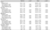
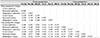
Mean central ST was 486.68 ± 25.15 µm, 535.16 ± 48.13 µm, 515.98 ± 44.07 µm, and 502.28 ± 34.87 µm preoperatively, and at 3 days, 1 week, and 1 month postoperatively, respectively (p < 0.001 for all). ST showed significant thickening in all three zones from 3 days to 1 month postoperatively (p < 0.001 for all). Mean central, paracentral, and midperipheral ET was 52.13 ± 3.41 µm, 50.42 ± 2.97 µm, 49.12 ± 3.05 µm at preoperatively and 51.03 ± 3.63 µm, 48.96 ± 3.62 µm, 47.67 ± 3.81 µm at 1 month postoperatively, respectively (p = 0.061, 0.006, 0.001, respectively), while there were no signficant changes in all three zones at 3 and 7 days postoperatively. Changes in ET and ST were prominent at the superotemporal incision site.
Conclusions
After scleral tunnel cataract surgery, corneal edema was observed in the stroma immediately after surgery. There was no significant change at early times after surgery in the epithelium, and there was a decrease in the peripheral cornea at 1 month postoperatively. The change in ET was considered a compensatory change due to stromal edema and appeared between 1 week to 1 month postoperatively.
Figures and Tables
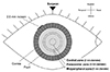 | Figure 1Schematic diagram of the cornea showing position of surgeon and analyzed area by RTVue. This diagram shows position of surgeon, incision site and analyzed areas on the cornea.
|
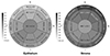 | Figure 2Preoperative corneal thickness. The preoperative epithelial thickness was thinner in the superior than inferior, but stromal thickness was thicker in the superior than inferior. S = superior; N = nasal; T = temporal; I = inferior.
|
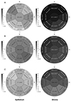 | Figure 3Postoperative changes of corneal thickness. (A) 3 days postoperative. (B) 1 week postoperative. (C) 1 month postoperative. *p < 0.05; **p < 0.01 in repeated measure analysis of variance (ANOVA) with post-hoc comparison compared to preoperative thickness. S = superior; N = nasal; T = temporal; I = inferior.
|
 | Figure 4Epithelial thickness changes among nuclear grade groups. (A) Central. (B) Paracentral. (C) Midperiphery. (D) Topographic variability. Pre-op = preoperative.
|
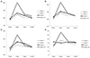 | Figure 5Stromal thickness changes among nuclear grade groups. (A) Central. (B) Paracentral. (C) Midperiphery. (D) Topographic variability. Pre-op = preoperative.
|

Notes
References
1. Hollows F, Moran D. Cataract--the ultraviolet risk factor. Lancet. 1981; 2:1249–1250.
2. Javitt JC, Wang F, West SK. Blindness due to cataract: epidemiology and prevention. Annu Rev Public Health. 1996; 17:159–177.
3. Asbell PA, Dualan I, Mindel J, et al. Age-related cataract. Lancet. 2005; 365:599–609.
4. Javitt JC, Steinert RF. Cataract extraction with multifocal intraocular lens implantation: a multinational clinical trial evaluating clinical, functional, and quality-of-life outcomes. Ophthalmology. 2000; 107:2040–2048.
5. Walkow T, Anders N, Klebe S. Endothelial cell loss after phacoemulsification: relation to preoperative and intraoperative parameters. J Cataract Refract Surg. 2000; 26:727–732.
6. Bourne RR, Minassian DC, Dart JK, et al. Effect of cataract surgery on the corneal endothelium: modern phacoemulsification compared with extracapsular cataract surgery. Ophthalmology. 2004; 111:679–685.
7. Zetterström C, Laurell CG. Comparison of endothelial cell loss and phacoemulsification energy during endocapsular phacoemulsification surgery. J Cataract Refract Surg. 1995; 21:55–58.
8. Clark A, Morlet N, Ng JQ, et al. Whole population trends in complications of cataract surgery over 22 years in Western Australia. Ophthalmology. 2011; 118:1055–1061.
9. Kanellopoulos AJ, Asimellis G. Corneal epithelial remodeling following cataract surgery: three-dimensional investigation with anterior-segment optical coherence tomography. J Refract Surg. 2014; 30:348–353.
10. Stein JD, Grossman DS, Mundy KM, et al. Severe adverse events after cataract surgery among medicare beneficiaries. Ophthalmology. 2011; 118:1716–1723.
11. Behndig A, Lundberg B. Transient corneal edema after phacoemulsification: comparison of 3 viscoelastic regimens. J Cataract Refract Surg. 2002; 28:1551–1556.
12. Mathew PT, David S, Thomas N. Endothelial cell loss and central corneal thickness in patients with and without diabetes after manual small incision cataract surgery. Cornea. 2011; 30:424–428.
13. Calabuig-Goena M, López-Miguel A, Marqués-Fernández V, et al. Early changes in corneal epithelial thickness after cataract surgery--pilot study. Curr Eye Res. 2016; 41:311–317.
14. Lundberg B, Jonsson M, Behndig A. Postoperative corneal swelling correlates strongly to corneal endothelial cell loss after phacoemulsification cataract surgery. Am J Ophthalmol. 2005; 139:1035–1041.
15. Zheng T, Yang J, Xu J, et al. Near-term analysis of corneal epithelial thickness after cataract surgery and its correlation with epithelial cell changes and visual acuity. J Cataract Refract Surg. 2016; 42:420–426.
16. Reinstein DZ, Archer T. Combined Artemis very high-frequency digital ultrasound-assisted transepithelial phototherapeutic keratectomy and wavefront-guided treatment following multiple corneal refractive procedures. J Cataract Refract Surg. 2006; 32:1870–1876.
17. Ventura AC, Wälti R, Böhnke M. Corneal thickness and endothelial density before and after cataract surgery. Br J Ophthalmol. 2001; 85:18–20.
18. Aribaba OT, Adenekan OA, Onakoya AO, et al. Central corneal thickness changes following manual small incision cataract surgery. Clin Ophthalmol. 2015; 9:151–155.
19. Şimşek A, Bilgin B, Çapkin M, et al. Evaluation of anterior segment parameter changes using the Sirius after uneventful phacoemulsification. Korean J Ophthalmol. 2016; 30:251–257.
20. Sanchis-Gimeno JA, Lieó-Pérez A, Alonso L, et al. Corneal endothelial cell density decreases with age in emmetropic eyes. Histol Histopathol. 2005; 20:423–427.
21. Siu AW, Herse PR. The effect of age on the edema response of the central and mid‐peripheral cornea. Acta Ophthalmol (Copenh). 1993; 71:57–61.
22. Cooper BA, Holekamp NM, Bohigian G, Thompson PA. Case-control study of endophthalmitis after cataract surgery comparing scleral tunnel and clear corneal wounds. Am J Ophthalmol. 2003; 136:300–305.
23. Ernest PH, Lavery KT, Kiessling LA. Relative strength of scleral corneal and clear corneal incisions constructed in cadaver eyes. J Cataract Refract Surg. 1994; 20:626–629.
24. Bolz M, Sacu S, Drexler W, Findl O. Local corneal thickness changes after small-incision cataract surgery. J Cataract Refract Surg. 2006; 32:1667–1671.
25. Reinstein DZ, Archer TJ, Gobbe M. Change in epithelial thickness profile 24 hours and longitudinally for 1 year after myopic LASIK: three-dimensional display with Artemis very high-frequency digital ultrasound. J Refract Surg. 2012; 28:195–201.
26. Reinstein DZ, Archer TJ, Gobbe M, et al. Epithelial thickness after hyperopic LASIK: three-dimensional display with Artemis very high-frequency digital ultrasound. J Refract Surg. 2010; 26:555–564.
27. Ytteborg J, Dohlman CH. Corneal edema and intraocular pressure. II. Clinical results. Arch Ophthalmol. 1965; 74:477–484.
28. Borderie VM, Baudrimont M, Vallée A, et al. Corneal endothelial cell apoptosis in patients with Fuchs' dystrophy. Invest Ophthalmol Vis Sci. 2000; 41:2501–2505.
29. Liesegang TJ. Physiologic changes of the cornea with contact lens wear. CLAO J. 2002; 28:12–27.
30. Morikubo S, Takamura Y, Kubo E, et al. Corneal changes after small-incision cataract surgery in patients with diabetes mellitus. Arch Ophthalmol. 2004; 122:966–969.
31. Mahdy MA, Eid MZ, Mohammed MA, et al. Relationship between endothelial cell loss and microcoaxial phacoemulsification parameters in noncomplicated cataract surgery. Clin Ophthalmol. 2012; 6:503–510.
32. Prasher P. Acute corneal melt associated with topical bromfenac use. Eye Contact Lens. 2012; 38:260–262.
 XML Download