

 PDF
PDF ePub
ePub Citation
 Print
Print


Abstract
Purpose
We report a rare case of bilateral macular infarction as an ocular presenting sign of primary antiphospholipid syndrome.
Case summary
A 29-year-old woman who had undergone a cesarean section for chorioamnionitis in the department of Obsterics was referred to the department of ophthalmology for bilateral visual loss. At examination, best-corrected visual acuity (BCVA) of the right eye was counting fingers, and for the left was 0.05. Fundus examination revealed extensive macular edema and cotton-wool spots in both eyes. We performed hematologic tests including thrombophilia examination. Antinuclear antibody and rheumatoid factor were negative but lupus anticoagulant presented high titers on two occasions 12 weeks apart. She was administered sub-Tenon's injections of triamcinolone acetonide 50 mg/week in both eyes under the diagnosis of bilateral macular arteriolar occlusion in primary antiphospholipid syndrome. Her BCVA remained 0.025 in her right eye and improved to 0.125 in her left eye.
Figures and Tables
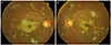 | Figure 1Fundus photographs showed confluent macular cotton wool spots and retinal ischemic edema presenting like cheery-red spots and intraretinal hemorrhages. (A) Right eye. (B) Left eye.
|
References
1. Behbehani R, Sergott RC, Savino PJ. The antiphospholipid antibody syndrome: diagnostic aspects. Curr Opin Ophthalmol. 2004; 15:483–485.
2. Cobo-Soriano R, Sánchez-Ramón S, Aparicio MJ, et al. Antiphospholipid antibodies and retinal thrombosis in patients without risk factors: a prospective case-control study. Am J Ophthalmol. 1999; 128:725–732.
3. Franchini M, Veneri D. The antiphospholipid syndrome. Hematology. 2005; 10:265–269.
4. Miserocchi E, Baltatzis S, Foster CS. Ocular features associated with anticardiolipin antibodies: a descriptive study. Am J Ophthalmol. 2001; 131:451–456.
5. Levine JS, Branch DW, Rauch J. The antiphospholipid syndrome. N Engl J Med. 2002; 346:752–763.
6. Montehermoso A, Cervera R, Font J, et al. Association of antiphospholipid antibodies with retinal vascular disease in systemic lupus erythematosus. Semin Arthritis Rheum. 1999; 28:326–332.
7. Kleiner RC, Najarian LV, Schatten S, et al. Vaso-occlusive retinopathy associated with antiphospholipid antibodies (lupus anticoagulant retinopathy). Ophthalmology. 1989; 96:896–904.
8. Nag TC, Wadhwa S. Histopathological changes in the eyes in systemic lupus erythematosus: an electron microscope and immunohistochemical study. Histol Histopathol. 2005; 20:373–382.
9. Shein J, Shukla D, Reddy S, et al. Macular infarction as a presenting sign of systemic lupus erythematosus. Retin Cases Brief Rep. 2008; 2:55–60.
10. Cooper BA, Shah GK, Grand MG. Purtscher's-like retinopathy in a patient with systemic lupus erythematosus. Ophthalmic Surg Lasers Imaging. 2004; 35:438–439.
11. Yoon CK, Park JH, Yu HG. Retinopathy associated with systemic lupus erythematosus. J Korean Ophthalmol Soc. 2009; 50:1215–1220.
12. Kim IT, Na SC, Lee KJ. Vascular occlusions associated with antiphospholipid antibodies in systemic lupus erythematosus. J Korean Ophthalmol Soc. 2000; 41:427–432.
13. Gass JD. A fluorescein angiographic study of macular dysfunction secondary to retinal vascular disease: V. Retinal telangiectasis. Arch Ophthalmol. 1968; 80:592–605.
14. Lahey JM, Tunç M, Kearney J, et al. Laboratory evaluation of hypercoagulable states in patients with central retinal vein occlusion who are less than 56 years of age. Ophthalmology. 2002; 109:126–131.
15. Castañón C, Amigo MC, Bañales JL, et al. Ocular vaso-occlusive disease in primary antiphospholipid syndrome. Ophthalmology. 1995; 102:256–262.
16. Al-Abdulla NA, Thompson JT, LaBorwit SE. Simultaneous bilateral central retinal vein occlusion associated with anticardiolipin antibodies in leukemia. Am J Ophthalmol. 2001; 132:266–268.
17. Kim SG, Kim YY, Song GG, et al. Primary antiphospholipid syndrome associated with non-ischemic central retinal vein occlusion. J Korean Ophthalmol Soc. 1995; 36:525–530.
18. Au A, O'Day J. Review of severe vaso-occlusive retinopathy in systemic lupus erythematosus and the antiphospholipid syndrome: associations, visual outcomes, complications and treatment. Clin Experiment Ophthalmol. 2004; 32:87–100.
19. Hong-Kee N, Mei-Fong C, Azhany Y, Zunaina E. Antiphospholipid syndrome in lupus retinopathy. Clin Ophthalmol. 2014; 8:2359–2363.
20. Hu CL, Peng KL. Bilateral macular infarction as an ocular manifestation of systemic lupus erythematous (SLE). Clin Ophthalmol. 2014; 8:1845–1848.
 XML Download