

 PDF
PDF ePub
ePub Citation
 Print
Print


Abstract
Purpose
To compare the clinical outcome of combined small incision lenticule extraction and collagen cross-linking (SMILE Xtra) with SMILE.
Methods
This study included 30 eyes from 15 patients who had undergone SMILE Xtra and a random sample of 30 eyes from 15 patients receiving SMILE alone during the same period. We obtained the following parameters from all patients: uncorrected (UDVA) and corrected distance visual acuity (CDVA), spherical equivalent (SE), efficacy and safety index, and corneal high-or-der aberrations.
Results
The SMILE Xtra group had higher preoperative SE and thinner central corneal and residual stromal bed thickness and optic zone diameter compared to the control group (p < 0.001). At 6 months, there was no significant difference in UDVA or CDVA between the two groups. The efficacy indices were 0.97 ± 0.16 and 1.05 ± 0.17 in the SMILE Xtra and control groups, re-spectively (p = 0.044), and there was no significant difference in safety index between the two groups during the follow-up period. Total corneal high-order aberrations numbered 2.59 ± 0.56 and 2.02 ± 0.41 in the SMILE Xtra and control groups, respectively (p <0.001), and there was significant increase in spherical aberration and horizontal corneal aberration in both groups compared to preoperative results. Corneal haze was observed in 20% of eyes in the SMILE Xtra group, and no complication such as corneal ectasia was observed during the follow-up period.
Go to : 
References
1. Binder PS. Ectasia after laser in situ keratomileusis. J Cataract Refract Surg. 2003; 29:2419–29.

2. Alió JL, Muftuoglu O, Ortiz D, et al. Ten-year follow-up of laser in situ keratomileusis for high myopia. Am J Ophthalmol. 2008; 145:55–64.
3. Raiskup-Wolf F, Hoyer A, Spoerl E, Pillunat LE. Collagen abdominal with riboflavin and ultraviolet-A light in keratoconus: long-term results. J Cataract Refract Surg. 2008; 34:796–801.
4. Hafezi F, Kanellopoulos J, Wiltfang R, Seiler T. Corneal collagen crosslinking with riboflavin and ultraviolet A to treat induced abdominal after laser in situ keratomileusis. J Cataract Refract Surg. 2007; 33:2035–40.
5. Kanellopoulos AJ, Binder PS. Collagen abdominal (CCL) with sequential topography-guided PRK: a temporizing alternative for keratoconus to penetrating keratoplasty. Cornea. 2007; 26:891–5.
6. Kanellopoulos AJ, Pamel GJ. Review of current indications for combined very high fluence collagen abdominal and laser in situ keratomileusis surgery. Indian J Ophthalmol. 2013; 61:430–2.
7. Shah R, Shah S, Sengupta S. Results of small incision lenticule abdominal: All-in-one femtosecond laser refractive surgery. J Cataract Refract Surg. 2011; 37:127–37.
8. Wang Y, Cui C, Li Z, et al. Corneal ectasia 6.5 months after small-incision lenticule extraction. J Cataract Refract Surg. 2015; 41:1100–6.
9. Mattila JS, Holopainen JM. Bilateral ectasia after femtosecond abdominal-assisted small incision lenticule extraction (SMILE). J Refract Surg. 2016; 32:497–500.
10. Ganesh S, Brar S. Clinical outcomes of small incision lenticule abdominal with accelerated abdominal (ReLEx SMILE Xtra) in abdominals with thin corneas and borderline topography. J Ophthalmol. 2015; 2015:263412.
11. Randleman JB, Woodward M, Lynn MJ, Stulting RD. Risk abdominal for ectasia after corneal refractive surgery. Ophthalmology. 2008; 115:37–50.
12. Nakamura K, Kurosaka D, Bissen-Miyajima H, Tsubota K. Intact corneal epithelium is essential for the prevention of stromal haze after laser assisted in situ keratomileusis. Br J Ophthalmol. 2001; 85:209–13.
13. Kobashi H, Kamiya K, Igarashi A, et al. Two-years results of small-incision lenticule extraction and wavefront-guided laser in situ keratomileusis for myopia. Acta Ophthalmol 2017 Jun 20. doi:. DOI: 10.1111/aos.13470. [Epub ahead of print].
14. Cai WT, Liu QY, Ren CD, et al. Dry eye and corneal sensitivity abdominal small incision lenticule extraction and femtosecond laser-abdominal in situ keratomileusis: a Meta-analysis. Int J Ophthalmol. 2017; 10:632–8.
15. Jin Y, Wang Y, Xu L, et al. Comparison of the optical quality abdominal small incision lenticule extraction and femtosecond laser LASIK. J Ophthalmol. 2016; 2016:2507973.
16. Wu D, Wang Y, Zhang L, et al. Corneal biomechanical effects: small-incision lenticule extraction versus femtosecond laser-abdominal laser in situ keratomileusis. J Cataract Refract Surg. 2014; 40:954–62.
17. Wang D, Liu M, Chen Y, et al. Differences in the corneal abdominal changes after SMILE and LASIK. J Refract Surg. 2014; 30:702–7.
18. Pedersen IB, Bak-Nielsen S, Vestergaard AH, et al. Corneal abdominal properties after LASIK, ReLEx flex, and ReLEx smile by Scheimpflug-based dynamic tonometry. Graefes Arch Clin Exp Ophthalmol. 2014; 252:1329–35.
19. Sefat SM, Wiltfang R, Bechmann M, et al. Evaluation of changes in human corneas after femtosecond laser-assisted LASIK and small-incision lenticule extraction (SMILE) using non-contact abdominal and ultra-high-speed camera (Corvis ST). Curr Eye Res. 2016; 41:917–22.
20. Sachdev G, Sachdev MS, Sachdev R, Gupta H. Unilateral corneal ectasia following small-incision lenticule extraction. J Cataract Refract Surg. 2015; 41:2014–8.
21. Tomita M, Yoshida Y, Yamamoto Y, et al. In vivo confocal laser abdominal of morphologic changes after simultaneous LASIK and accelerated collagen crosslinking for myopia: one-year results. J Cataract Refract Surg. 2014; 40:981–90.
22. Kanellopoulos AJ, Asimellis G, Karabatsas C. Comparison of abdominal higher fluence corneal abdominal to control, in abdominal LASIK, one year results. Clin Ophthalmol. 2014; 8:2373–81.
23. Ng AL, Chan TC, Cheng GP, et al. Comparison of the early clinical outcomes between combined small-incision lenticule extraction and collagen abdominal versus SMILE for myopia. J Ophthalmol. 2016; 2016:2672980.
24. Wu W, Wang Y, Zhang H, et al. One-year visual outcome of small incision lenticule extraction (SMILE) surgery in high myopic eyes: retrospective cohort study. BMJ Open. 2016; 6:e010993.
25. Celik HU, Alagöz N, Yildirim Y, et al. Accelerated corneal abdominal concurrent with laser in situ keratomileusis. J Cataract Refract Surg. 2012; 38:1424–31.
26. Kanellopoulos AJ, Asimellis G. Epithelial remodeling after abdominal laser-assisted high myopic LASIK: comparison of stand-alone with LASIK combined with prophylactic high-fluence abdominal. Cornea. 2014; 33:463–9.
27. Alhayek A, Lu PR. Corneal collagen crosslinking in keratoconus and other eye disease. Int J Ophthalmol. 2015; 8:407–18.
28. Greenstein SA, Fry KL, Bhatt J, Hersh PS. Natural history of abdominal haze after collagen crosslinking for keratoconus and corneal ectasia: Scheimpflug and biomicroscopic analysis. J Cataract Refract Surg. 2010; 36:2105–14.
29. Koppen C, Vryghem JC, Gobin L, Tassignon MJ. Keratitis and abdominal scarring after UVA/riboflavin abdominal for keratoconus. J Refract Surg. 2009; 25:S819–23.
30. Kymionis GD, Tsoulnaras KI, Grentzelos MA, et al. Corneal abdominal demarcation line after standard and high-intensity collagen crosslinking determined with anterior segment optical coherence tomography. J Cataract Refract Surg. 2014; 40:736–40.
31. Ağ ca A, Demirok A, Cankaya K, et al. Comparison of visual acuity and higher-order aberrations after femtosecond lenticule extraction and small-incision lenticule extraction. Cont Lens Anterior Eye. 2014; 37:292–6.
32. Jin HY, Wan T, Wu F, Yao K. Comparison of visual results and higher-order aberrations after small incision lenticule extraction (SMILE): high myopia vs. mild to moderate myopia. BMC Ophthalmol. 2017; 17:118.
Go to :
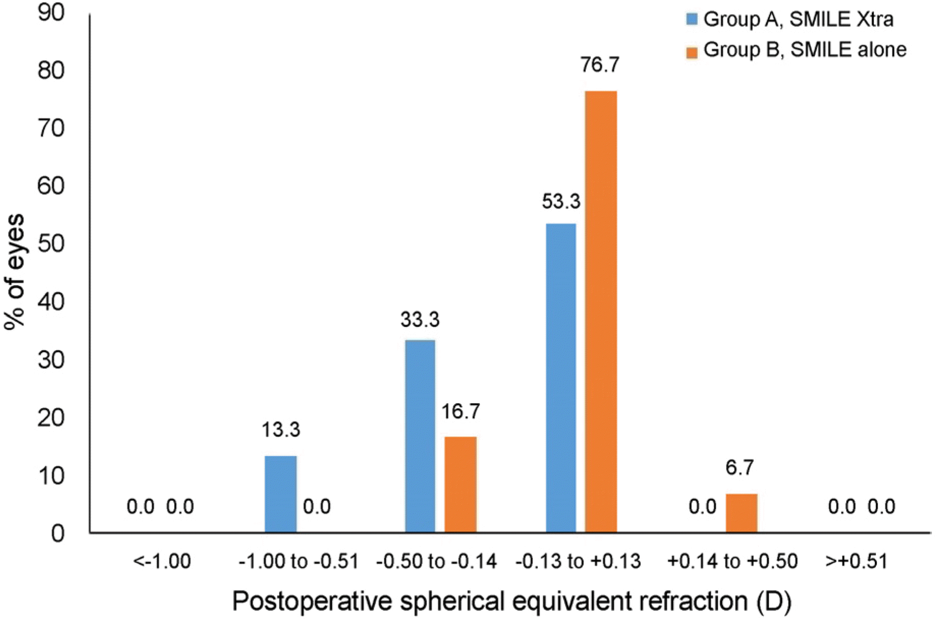 | Figure 1.Refractive outcomes for small incision lenticlue extraction (SMILE) and SMILE Xtra at 6 months postoperatively. Percentages of eyes within different diopter ranges of the intended correction in spherical equivalent refraction in both groups. |
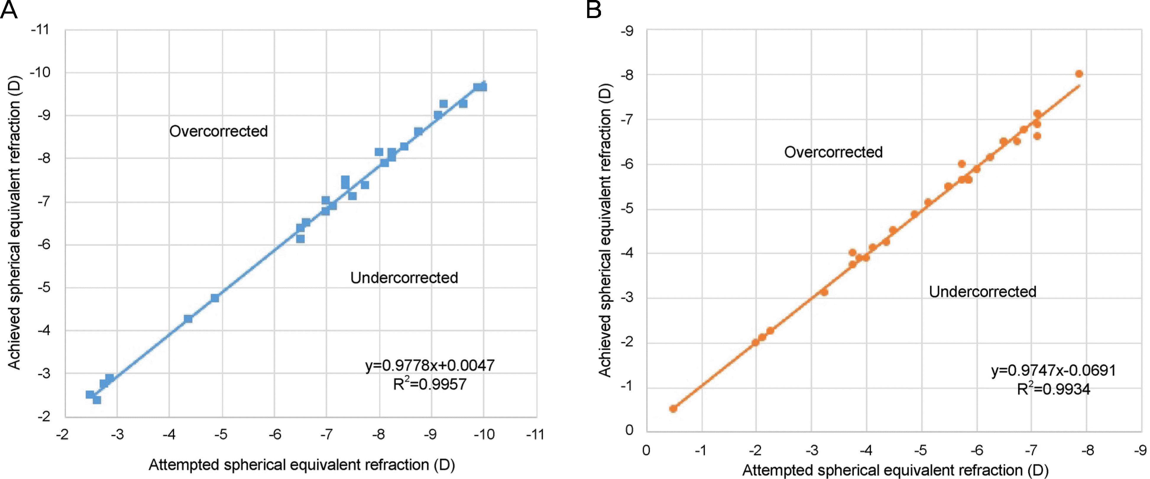 | Figure 2.Predictability of spherical equivalent correction at 6 months, in (A) SMILE Xtra group and (B) SMILE alone group. Both groups showed a high level of refractive predictability. SMILE = small incision lenticlue extraction. |
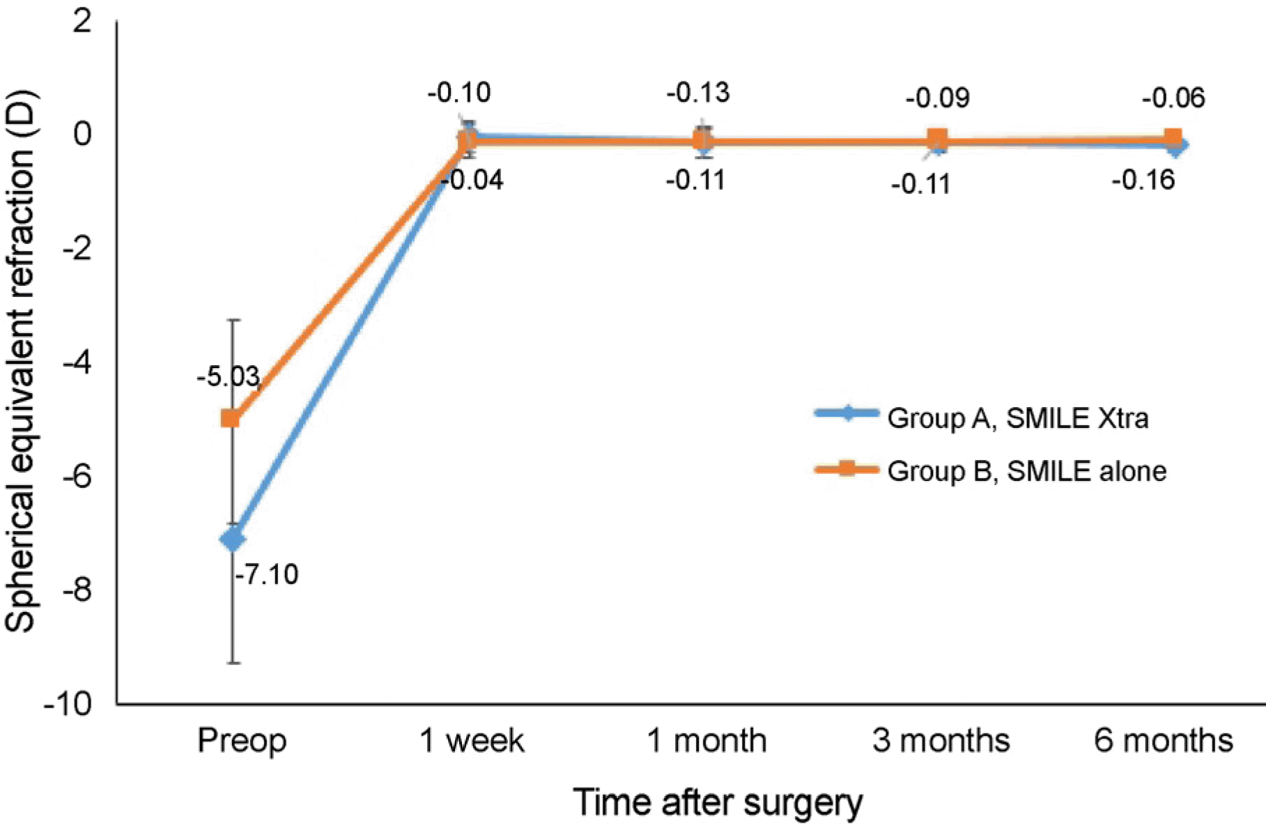 | Figure 3.Stability of spherical equivalent refraction in both groups after surgery. In group B, spherical equivalent was stable for 6 months after the surgery, but over time, spher-ical equivalent was incrased in group A. |
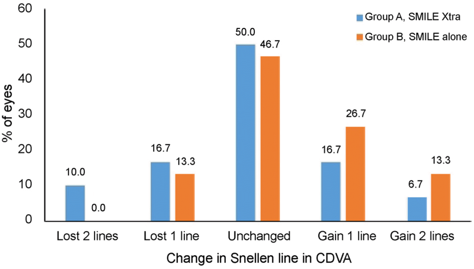 | Figure 4.Change in corrected distance visual acuity (CDVA). Gain and loss in Snellen lines of both groups 6 months postoperatively. |
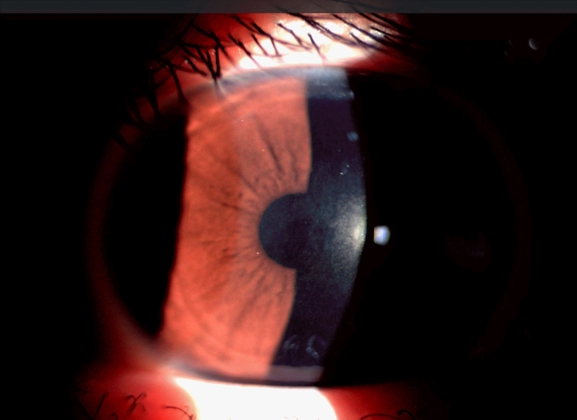 | Figure 5.Anterior segment photograph of the patient who has Grade 0.5 corneal haze after SMILE Xtra. The cornea has faint hazziness at center, but the patient dose not complained any clinical symptoms. SMILE = small incision lenticlue extraction. |
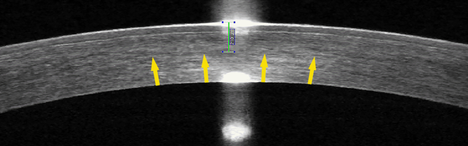 | Figure 6.Anterior segment optical coherence tomography 6 months postoperatively. Hyper-reflection line is seen faintly (arrows). The depth of demarcation line was 210 μ m (green line). |
Table 1.
Preoperative demographics
| SMILE Xtra | SMILE alone | p-value* | |
|---|---|---|---|
| Age (years) | 27.07 | 27.47 | N/A |
| Gender (male) (n, %) | 9 (60) | 5 (33.3) | N/A |
| Sphere (diopter) | –6.44 ± 2.07 | –4.42 ± 1.67 | <0.001 |
| Cylinder (diopter) | –1.24 ± 0.85 | –1.22 ± 0.84 | 0.537 |
| Spherical equivalent (diopter) | –7.06 ± 2.17 | –5.03 ± 1.78 | <0.001 |
| Flat sim K (diopter) | 42.76 ± 1.35 | 42.25 ± 1.13 | 0.247 |
| Steep sim K (diopter) | 44.43 ± 1.90 | 44.06 ± 1.43 | 0.214 |
| Central corneal thickness (μ m) | 515.03 ± 23.79 | 544.87 ± 26.46 | <0.001 |
| Planned residual stromal bed (μ m) | 281.43 ± 24.29 | 328.27 ± 33.69 | <0.001 |
| Scotopic pupil size (mm) | 6.24 ± 0.71 | 6.39 ± 0.59 | 0.720 |
| Intended optic zone (mm) | 6.18 ± 0.25 | 6.47 ± 0.14 | <0.001 |
Table 2.
Comparison of uncorrected and corrected distance visual acuity of both groups
|
POD |
|||||
|---|---|---|---|---|---|
| 1 week | 1 month | 3 months | 6 months | ||
| UDVA (logMAR) | SMILE Xtra | –0.03 ± 0.05 | –0.05 ± 0.07 | –0.06 ± 0.05 | –0.06 ± 0.04 |
| SMILE alone | –0.06 ± 0.06 | –0.09 ± 0.05 | –0.08 ± 0.04 | –0.09 ± 0.05 | |
| p-value* | 0.091 | 0.219 | 0.217 | 0.095 | |
| CDVA (logMAR) | SMILE Xtra | –0.04 ± 0.05 | –0.06 ± 0.06 | –0.08 ± 0.05 | –0.08 ± 0.06 |
| SMILE alone | –0.05 ± 0.05 | –0.09 ± 0.04 | –0.10 ± 0.05 | –0.10 ± 0.05 | |
| p-value* | 0.874 | 0.171 | 0.344 | 0.146 | |
Table 3.
The efficacy indices (postoperative UDVA/preoperative CDVA) and the safety indices (postoperative CDVA/preoperative CDVA) of both groups
|
POD |
|||
|---|---|---|---|
| 3 months | 6 months | ||
| Efficacy index | SMILE Xtra | 0.97 ± 0.13 | 0.97 ± 0.16 |
| SMILE alone | 1.01 ± 0.14 | 1.05 ± 0.17 | |
| p-value* | 0.249 | 0.044 | |
| Safety index | SMILE Xtra | 1.01 ± 0.19 | 1.01 ± 0.21 |
| SMILE alone | 1.08 ± 0.17 | 1.09 ± 0.20 | |
| p-value* | 0.180 | 0.102 | |
Table 4.
Preoperative and 6-month postoperative corneal aberrations measured by Galilei®
|
Preoperative |
6-month postoperative |
Difference pre/post p-value† |
||||||
|---|---|---|---|---|---|---|---|---|
| SMILE Xtra | SMILE alone | p-value* | SMILE Xtra | SMILE alone | p-value* | SMILE Xtra | SMILE alone | |
| RMS total | 1.84 ± 0.76 | 2.1 ± 0.9 | 0.287 | 2.59 ± 0.56 | 2.02 ± 0.41 | <0.001 | <0.001 | 0.307 |
| Vertical Coma, Z3−1 | –0.01 ± 0.15 | –0.06 ± 0.34 | 0.739 | 0.02 ± 0.32 | –0.02 ± 0.31 | 0.889 | 0.447 | 0.421 |
| Horizontal Coma, Z31 | 0.01 ± 0.19 | 0.05 ± 0.38 | 0.371 | –0.22 ± 0.3 | –0.16 ± 0.32 | 0.409 | <0.001 | <0.001 |
| Vertical Trefoil, Z3−3 | –0.13 ± 0.12 | –0.03 ± 0.22 | 0.133 | –0.07 ± 0.21 | –0.08 ± 0.19 | 0.827 | 0.073 | 0.352 |
| Oblique Trefoil, Z33 | –0.03 ± 0.19 | –0.07 ± 0.34 | 0.222 | –0.05 ± 0.23 | –0.02 ± 0.15 | 0.161 | 0.476 | 0.280 |
| Spherical aberration, Z40 | 0.22 ± 0.1 | 0.22 ± 0.1 | 0.610 | 0.52 ± 0.2 | 0.32 ± 0.12 | <0.001 | <0.001 | 0.001 |
 XML Download