Journal List > J Korean Ophthalmol Soc > v.58(10) > 1010643

Kim, Seo, and Yoon: A Retrospective Analysis of Granulomatosis with Polyangiitis with Ocular Manifestations

Abstract

Purpose

To analyze the treatment and prognosis of patients from our tertiary medical center with ocular and orbital involvement of granulomatosis with polyangiitis.

Methods

A retrospective analysis of the medical records of patients diagnosed with granulomatosis with polyangiitis that visited our single tertiary referral center from July 2008 to September 2014 was performed.

Results

A total of 51 patients diagnosed with granulomatosis with polyangiitis visited our center, and 21 of those patients had received an ophthalmologic examination. Of these, 9 patients (4 males, 5 females) had symptoms of the eye and orbit, and the clinical presentations were as follows: episcleritis, scleritis, marginal keratitis, orbital inflammation, orbital abscess, retinal vasculitis, and nasolacrimal duct obstruction. The patients each received treatments according to clinical presentation with topical, oral, or intravenous steroids or immunomodulatory agents such as cyclophosphamide. Nasolacrimal duct obstruction was treated with surgery in some cases. After an average follow-up period of 58 ± 30 months, all patients showed clinical improvement of their ocular and orbital involvement of granulomatosis with polyangiitis.

Conclusions

Granulomatosis with polyangiitis is a relatively rare disease that sometimes has ocular or orbital involvement and can lead to blindness. Therefore, when ocular symptoms and signs present without a definitive cause, granulomatosis with polyangiitis must be ruled out, and appropriate treatment is needed. However, there are few published reports on the clinical presentation and prognosis of ocular and orbital involvement of granulomatosis with polyangiitis in Asians. This study showed that the incidence of ocular and orbital involvement in granulomatosis with polyangiitis was lower than previous reports.

References

1. Falk RJ, Gross WL, Guillevin L, et al. Granulomatosis with polyangiitis (Wegener's): an alternative name for Wegener's granulomatosis. Arthritis Rheum. 2011; 63:863–4.
crossref
2. Fahey JL, Leonard E, Churg J, Godman G. Wegener's granulomatosis. Am J Med. 1954; 17:168–79.
crossref
3. Leavitt RY, Fauci AS, Bloch DA, et al. The American College of Rheumatology 1990 criteria for the classification of Wegener's granulomatosis. Arthritis Rheum. 1990; 33:1101–7.
crossref
4. Jennette JC, Falk RJ, Andrassy K, et al. Nomenclature of systemic vasculitides. Proposal of an international consensus conference. Arthritis Rheum. 1994; 37:187–92.
5. Savige J, Dimech W, Fritzler M, et al. Addendum to the International Consensus Statement on testing and reporting of antineutrophil abdominal antibodies. Quality control guidelines, comments, and abdominal for testing in other autoimmune diseases. Am J Clin Pathol. 2003; 120:312–8.
6. Rao JK, Weinberger M, Oddone EZ, et al. The role of abdominal cytoplasmic antibody (c-ANCA) testing in the abdominal of Wegener granulomatosis: a literature review and meta-analysis. Ann Intern Med. 1995; 123:925–32.
7. Harper SL, Letko E, Samson CM, et al. Wegener's granulomatosis: the relationship between ocular and systemic disease. J Rheumatol. 2001; 28:1025–32.
8. Reinhold-Keller E, Beuge N, Latza U, et al. An interdisciplinary approach to the care of patients with Wegener's granulomatosis: long-term outcome in 155 patients. Arthritis Rheum. 2000; 43:1021–32.
crossref
9. Hoffman GS, Kerr GS, Leavitt RY, et al. Wegener granulomatosis: an analysis of 158 patients. Ann Intern Med. 1992; 116:488–98.
crossref
10. Joshi L, Tanna A, McAdoo SP, et al. abdominal outcomes of abdominal therapy in ocular granulomatosis with polyangiitis: impact on localized and nonlocalized disease. Ophthalmology. 2015; 122:1262–8.
11. You C, Ma L, Lasave AF, Foster CS. Rituximab induction and maintenance treatment in patients with scleritis and granulomatosis with polyangiitis (Wegener's). Ocul Immunol Inflamm 2017 Jun 19. 1–8. doi:. DOI: 10.1080/09273948.2017.1327602. [Epub ahead of print].
crossref
12. Hellmich B, Flossmann O, Gross WL, et al. EULAR abdominal for conducting clinical studies and/or clinical trials in systemic vasculitis: focus on abdominal cytoplasm anti-body-associated vasculitis. Ann Rheum Dis. 2007; 66:605–17.
13. Hogan PC, O'Connell RM, Scollard S, et al. Biomarkers predict abdominal in granulomatosis with polyangiitis. J Biomark. 2014; 2014:596503.
14. Lee SJ, Kim SS, Kim WS. A case of Wegener's granulomatosis with retinal pigment epithelial detachment and subretinal hemorrhage. J Korean Ophthalmol Soc. 2005; 46:176–85.
15. Choi JS, Choi KY. Five cases of Wegener's granulomatosis with ocular manifestations. J Korean Ophthalmol Soc. 1986; 27:903–10.
16. Jabs DA, Mudun A, Dunn JP, Marsh MJ. Episcleritis and scleritis: clinical features and treatment results. Am J Ophthalmol. 2000; 130:469–76.
crossref
17. Bullen CL, Liesegang TJ, McDonald TJ, DeRemee RA. Ocular complications of Wegener's granulomatosis. Ophthalmology. 1983; 90:279–90.
crossref
18. Terrier B, Dechartres A, Deligny C, et al. Granulomatosis with abdominal according to geographic origin and ethnicity: clinical-biological presentation and outcome in a French population. Rheumatology. 2017; 56:445–50.
19. Sugiyama K, Sada KE, Kurosawa M, et al. Current status of the treatment of microscopic polyangiitis and granulomatosis with polyangiitis in Japan. Clin Exp Nephrol. 2013; 17:51–8.
crossref
20. Tsuchida Y, Shibuya M, Shoda H, et al. Characteristics of abdominal with polyangiitis patients in Japan. Mod Rheumatol. 2015; 25:219–23.
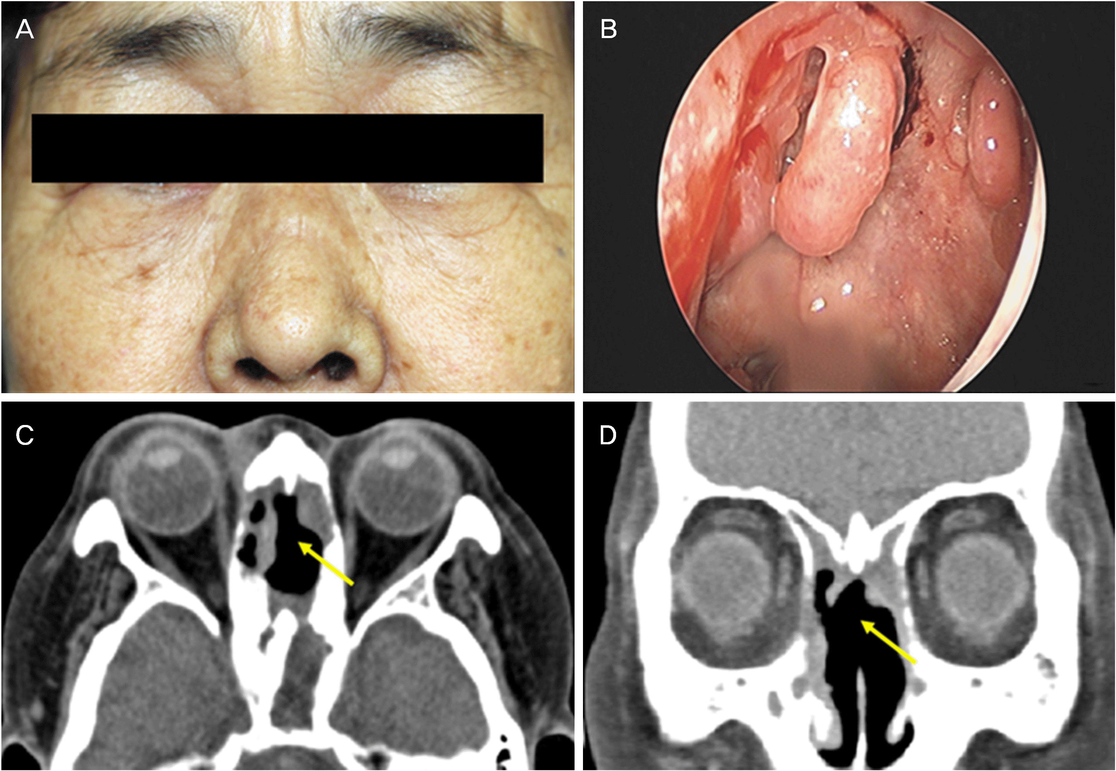
Figure 1.
Case 7. This 68-year-old woman presented with epiphora. She had a history of sinusitis and periorbital inflammation. (A) A photograph showing the collapse of the nasal bridge, also known as the ‘saddle nose’. (B) An endoscopic view of the right nasal cavity shows friable mucosa and easy bleeding. (C, D) The coronal and axial views of contrast computed tomography show chronic inflammatory changes in form of the destruction of the nasal septum and ethmoid sinus (yellow arrow).
jkos-58-1115f1.tif
Table 1.
Ophthalmic characteristics and the systemic involvement of patients with granulomatosis with polyangiitis
Patient No. Sex/Age Ophthalmic manifestation Chief complaint c-ANCA (PR3) p-ANCA (MPO) Other systemic involvement Other medical history
1 M/58 Episcleritis (OU) Congestion Positive (127.9) Negative Lung, Kidney Hypertension Prostate cancer
2 M/74 Scleritis (OU) Congestion, Ocular pain Positive (27.0) Positive (73.0) Lung, Kidney Hypertension
3 F/67 Retinal vasculitis (OD) Floater Positive Negative Lung, Sinus, Brain Bladder cancer
    Peripheral keratitis (OD)          
4 M/67 Orbital inflammation (OS) Periorbital swelling Positive (23.0) Negative Nose None
5 F/36 Dacryoadenitis (OU) Lid swelling Positive (72.7) Negative Lung Hyperthyroidism
6 M/32 Orbital inflammation (OU) NLD obstruction (OU) Tearing, Diplopia Positive (218.0) Positive (45.0) Lung, Sinus, Nose, Brain None
7 F/68 NLD obstruction (OU) Tearing Positive (82.8) Negative Sinus, Kidney Hypergammaglobulinemia
8 F/54 NLD obstruction (OU) Tearing Positive (70.0) Negative Sinus Hypertension
9 F/56 Orbital abscess (OD) Diplopia, Ocular pain, Lid swelling Negative Negative Lung, Kidney, Sinus None

All patients’ ophthalmic manifestations were improved after treatment. Patient 4, 8: Granulomatosis with Polyangiitis was diagnosed based on clinical findings of the eye and the orbit. c-ANCA = cytoplasmic antineutrophil cytoplasmic antibody; PR3 = proteinase 3; p-ANCA = perinuclear antineutrophil cytoplasmic antibody; MPO = myeloperoxidase; M = male; F = female; OU = oculus unitas; OD = oculus dexter; OS = oculus sinister; NLD = nasolacrimal duct.

Table 2.
Ocular and systemic treatment of patients with granulomatosis with polyangiitis
Patient No. Ophthalmic manifestation Treatment for eye Systemic treatment Pre VA (OD/OS) Post VA (OD/OS)
1 Episcleritis (OU) Topical steroid Steroid pulse (62.5 mg/day × 3 wks → 31.25 mg/day) UC/UC UC/UC
2 Scleritis (OU) Topical antibiotics PO steroid (5–10 mg/day) PO steroid (5–10 mg/day) Immunosuppressive agent (Azathioprine) 0.9/0.7 1.0/0.5
3 Retinal vasculitis (OD) Peripheral keratitis (OD) Retinal photocoagulation (OD) PO steroid (5–15 mg/day) PO steroid (5–15 mg/day) Immunosuppressive agent (Cyclophosphamide → Azathioprine), Lung resection, Transsphenoidal surgery 0.9/1.0 0.2/0.9
4 Orbital inflammation (OS) IV steroid (3.75 g) → PO steroid (25–40 mg) IV steroid (3.75 g) → PO steroid (25–40 mg) Immunosuppressive agent (Cyclophosphamide) 0.8/0.3 1.0/0.4
5 Dacryoadenitis (OU) Topical antibiotics and steroid PO steroid (5–30 mg/day) Steroid pulse (1 g) PO steroid (5–30 mg/day) Immunosuppressive agent (Cyclophosphamide) → d/c 0.8/0.8
6 Orbital inflammation (OU) NLD obstruction (OU) IV steroid (Medrol 12.5 g, Cortef 400 mg) Endoscopic DCR (OU) – The operation was done during remission Steroid pulse (4 times) PO steroid (5–60 mg/day) Immunosuppressive agent (Cyclophosphamide, Mycophenolate mofetil) 0.9/0.8 0.9/1.0
7 NLD obstruction (OU) Topical antibiotics and steroid Immunosuppressive agent (Cyclophosphamide pulse 10 times → Azathioprine 50–100 mg/day) PO steroid 2–10 mg/day 0.8/0.8 0.7/0.7
8 NLD obstruction (OU) Orbital cellulitis IV steroid (1 g) Endoscopic DCR (OU) – The 1st operation was done 2 weeks after IV steroid pulse therapy and the revision operation was done during remission IV antibiotics PO steroid (2.5–25 mg/day) Immunosuppressive agent (Methotrexate) 0.6/0.8 0.8/0.8
9 Orbital abscess (OD) Subperiosteal abscess drainage (OD) None (antibiotic therapy was initiated due to sign of the infection, but the follow up loss occurred.) 0.5/0.5 (near) 0.7/0.7

VA = visual acuity; OD = oculus dexter; OS = oculus sinister; OU = oculus unitas; wks = weeks; UC = uncheckable; PO = per oral; IV = intravenous; d/c = discontinue; NLD = nasolacrimal duct; DCR = dacryocystorhinostomy.

Table 3.
The characteristics of patients with granulomatosis with polyangiitis at the time of the diagnosis and the ophthalmic clinic visit
Patient No. Ophthalmic manifestation GPA Diagnosis date (symptom) First visit date (Ophthalmology) & GPA activity Remission after ocular symptom Last Post f/u lab f/u period (months)
1 Episcleritis (OU) Apr 07 2010 (ARDS → Lab review & Renal biopsy) Feb 22 2010, Active Unknown (transfer out) CRP 58
2 Scleritis (OU) 1999 (FUO → Renal biopsy, TBLB) Aug 24 2006, Remission Death c-ANCA (26.0), p-ANCA (172.0), CRP 60
3 Retinal vasculitis (OD) Peripheral keratitis (OD) 1993 (dyspnea, sputum → Abnormal Chest X-ray, ANCA+) Sep 04 2013, Active Death c-ANCA (negative), p-ANCA (negative), CRP 120
4 Orbital inflammation (OS)   May 08 2013, First diagnosis No c-ANCA (equivocal), p-ANCA (negative), CRP 19
5 Dacryoadenitis (OU) 2003 (nasal obstruction → Nasal septum biopsy, ANCA+) Nov 04 2008, Remission Unknown (transfer out) c-ANCA (negative), p-ANCA (negative), CRP 72
6 Orbital inflammation (OU) NLD obstruction (OU) 1998 (Nasal septum perforation → symptom & Lab review) Aug 04 2007, Remission Death c-ANCA (141.0), p-ANCA (31.0), CRP 72
7 NLD obstruction (OU) Aug 13 2008 (sinusitis → biopsy) Jan 02 2014, Active No c-ANCA (8.0), p-ANCA (negative), CRP 72
8 NLD obstruction (OU) Orbital cellulitis   Mar 17 2010, First diagnosis Yes c-ANCA (15.0), p-ANCA (negative), CRP 18
9 Orbital abscess (OD) Jun 25 2012 (Orbital pain → biopsy by ENT & symptom, Lab review) Jun 08 2012, Active Unknown (f/u loss) CRP 30

GPA = granulomatosis with polyangiitis; f/u = follow up; OU = oculus unitas; OD = oculus dexter; OS = oculus sinister; ARDS = acute respiratory distress syndrome; CRP = C reactive protein; FUO = fever of unknown origin; TBLB = transbronchial lung biopsy; c-ANCA = cytoplasmic antineutrophil cytoplasmic antibody; p-ANCA = perinuclear antineutrophil cytoplasmic antibody; NLD = nasolacrimal duct; ENT = ear nose throat.

TOOLS
Similar articles