Abstract
Purpose
To present a case of orbital inflammation and optic perineuritis preceding vesicular eruption in herpes zoster oph-thalmicus (HZO).
Case summary
An 84-year-old woman with a history of gall bladder cancer and hypertension complained of left periorbital eryth-ematous edema and discomfort. On examination, visual acuity was 20/25 bilaterally; no tenderness, proptosis or oph-thalmoplegia was observed. Pupils were equal, round, and reactive to light without relative afferent pupillary defects. Slit-lamp examination revealed severe conjunctival injection and chemosis without keratitis or uveitis. The remainder of the ocular exami-nation was unremarkable. Magnetic resonance imaging confirmed left-sided preseptal swelling with an enlarged left lacrimal gland, high signal intensity of the retrobulbar fat and optic nerve sheath. Systemic antibiotic therapy with steroids was started un-der a presumed diagnosis of idiopathic orbital inflammatory disease, but the clinical presentation was unresolved. After 2 days, vesicular lesions confined to the first division of the trigeminal nerve and pseudodendritic keratitis developed on the left side leading to a diagnosis of HZO. Treatment with acyclovir immediately resolved anterior segment inflammation and periorbital edema. While on therapy, visual acuity deteriorated to 20/125 and the pupil became dilated and unresponsive to light over a few days. All signs and symptoms of acute orbitopathy and postherpetic neuralgia had resolved 3 months later with the exception of pupil abnormality and visual acuity.
References
1. Liesegang TJ. Herpes zoster ophthalmicus: natural history, risk factors, clinical presentation, and morbidity. Ophthalmology 2008. 115; ((2 Suppl)):S3–12.
2. Shaikh S, Ta CN.Evaluation and management of herpes zoster ophthalmicus. Am Fam Physician. 2002; 66:1723–30.
3. Temnogorod J, Pointdujour-2 Lim, Mancini R. . Acute orbital syndrome in herpes zoster ophthalmicus: clinical features of 7 cases. Ophthal Plast Reconstr Surg. 2017; 33:173–7.

4. Volpe NJ, Shore JW. Orbital myositis associated with herpes zoster. Arch Ophthalmol. 1991; 109:471–2.

5. Kawasaki A, Borruat FX. An unusual presentation of herpes zoster ophthalmicus: orbital myositis preceding vesicular eruption. Am J Ophthalmol. 2003; 136:574–5.

6. Obata H, Yamagami S, Saito S. . A case of acute dacryoadenitis associated with herpes zoster ophthalmicus. Jpn J Ophthalmol. 2003; 47:107–9.

7. Tseng YH. Acute orbital myositis heralding herpes zoster oph-thalmicus: report of a case. Acta Neurol Taiwan. 2008; 17:47–9.
8. Kim HT, Moon SY, Lee KH. Acute orbital myositis before herpes zoster ophthalmicus. Korean J Anesthesiol. 2012; 62:295–6.

9. Bela C, Obéric A, Matet A. . Right acute dacryoadenitis shortly preceding ipsilateral herpes zoster ophthalmicus, a case report. Klin Monbl Augenheilkd. 2015; 232:497–9.

10. Patheja RS, Weaver T, Morris S. Unique case of orbital myositis and dacryoadenitis preceding the vesicular rash of herpes zoster ophthalmicus. Clin Exp Ophthalmol. 2016; 44:138–40.

11. Ota T, Yamazaki M, Toda Y. . A case of herpes zoster oph-thalmicus preceded one week by diplopia and ophthalmalgia. Rinsho Shinkeigaku. 2017; 57:163–7.

13. Ragozzino MW, Melton LJ 3rd, Kurland LT. . Population- based study of herpes zoster and its sequelae. Medicine (Baltimore). 1982; 61:310–6.
14. Vardy SJ, Rose GE. Orbital disease in herpes zoster ophthalmicus. Eye (Lond). 1994; 8((Pt 5)):577–9.

15. Han JB, Kim TG, Jin KH. Three cases of pupil abnormality in her-pes zoster ophthalmicus. J Korean Ophthalmol Soc. 2013; 54:1452–7.

16. Czyz CN, Bacon TS, Petrie TP. . Isolated, complete paralytic mydriasis secondary to herpes zoster ophthalmicus. Pract Neurol. 2013; 13:183–4.

Figure 1.
Clinical photographs. (A) Left periorbital eryth-ematous edema without tenderness, proptosis or ophthalmoplegia was observed along with conjunctival injection and chemosis at initial presentation. (B) Four days after initial presentation, vesic-ular eruptions involving the first division of the trigeminal nerve on left side developed.

Figure 2.
Magnetic resonance imaging findings. (A) Axial and (B, C) coronal sections showed left-sided preseptal swelling, enlarged lacrimal gland (arrow) and high signal intensity of the retrobulbar fat and linear signal change of the optic nerve sheath.

Figure 3.
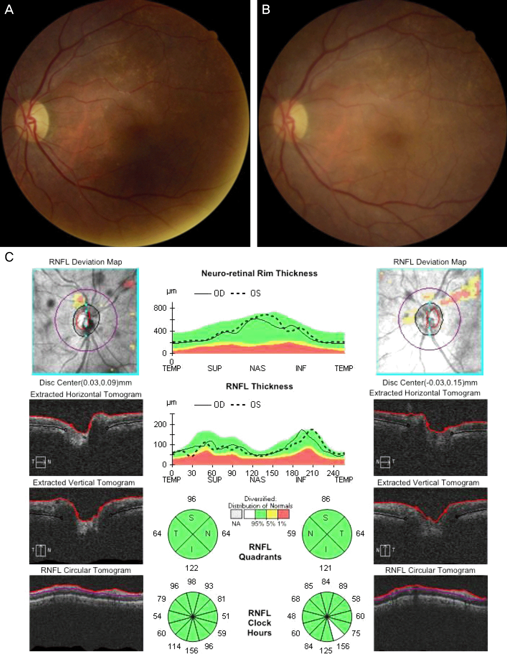
Fundus photographs and optical coherence tomography (OCT) image of the patient. (A) Fundus photograph of the left eye showed normal appearance of optic disc and posterior pole when the patient’s visual acuity deteriorated to 20/125 in spite of treatment. (B, C) Three months later, there was no optic disc pallor or significant peripapillary retinal nerve fiber layer defect on fundus photograph and OCT of the left eye. RNFL = retinal nerve fiber layer; OD = oculus dexter; OS = oculus sinister; TEMP = temporal; SUP = superior; NAS = nasal; INF = inferior; S = superior; N = nasal; I = inferior; T = temporal.


 PDF
PDF ePub
ePub Citation
 Print
Print


 XML Download