

 PDF
PDF ePub
ePub Citation
 Print
Print


Abstract
Purpose
In the present study, a case of double fovea artifact on spectral-domain optical coherence tomography (SD-OCT) was reported.
Case summary
A nine-year-old male presented with blurred vision of both eyes. His best corrected visual acuity (BCVA) was 20/20 in both eyes, and complete ophthalmologic evaluation including fundus examination and fundus photography revealed no abnormality in both eyes. He underwent SD-OCT imaging with the Cirrus HD-OCT. The Macular Cube 512 × 128 protocol of his right eye revealed an unusual pseudo-duplication of the fovea in the vertical meridian. The same protocol in his left eye also rendered a pseudo-duplication of two foveas in the vertical and horizontal meridians on the retinal thickness map. Re-examination with the same OCT system and protocol was performed two weeks later after the patient received counseling on fixation during the examination, and it revealed normal contour of the fovea in both eyes.
Conclusions
Double fovea artifact seen in SD-OCT is a rare artifact that can possibly lead to misdiagnosis and inappropriate clinical treatment. Since the artifact was resolved with better fixation of the patient, repeating the scan with better patient compliance is necessary when such an artifact is encountered.
Figures and Tables
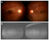 | Figure 1Fundus photography and autofluorescent photography of both eyes. Fundus photography (A), autofluorescent fundus photography (B) of a nine-years-old patient with double fovea artifact on HD-OCT scan.
|
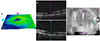 | Figure 2Cirrus HD-OCT of the right eye. (A) Retinal thickness map from the Macular Cube 512 × 128 volume scan. Two contiguous foveal depressions are present. (B) Horizontal, transfoveal B-scan of the right eye that demonstrates a normal foveal contour of one of two foveal depressions. (C) Vertical scan of the same eye. Note two foveal depressions. (D) Optical coherence tomography fundus image overlay (blue asterisk) on infrared photomicrograph (red asterisk) of the right eye. The abrupt transition is noted by white arrows. Note gray shadow distinguishable from other areas in the background.
|
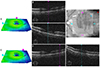 | Figure 3Cirrus HD-OCT of the left eye. (A) Retinal thickness map from the Macular Cube 512 × 128 volume scan on Cirrus HD-OCT. Three foveal depressions are noted. (B) Horizontal, transfoveal B-scan of the left eye that demonstrates a normal foveal contour. Foveal depression of the lateral side visible on retinal thickness map's horizontal meridian is presented not as a foveal pit, but as a normal retinal contour. (C) Vertical B-scan of the same eye presenting two foveal depressions. (D) Optical coherence tomography fundus image overlay (blue asterisk) on infrared photomicrograph (red asterisk) of the left eye. Some abrupt discontinuations of vessels are noted by white arrows. Note gray shadow distinguishable from other areas in the background. (E-G) Foveal duplication at the horizontal meridian to the anatomical fovea shown in retinal thickness map is not clearly presented in both horizontal and vertical scans. However, signal intensity at the site of foveal duplication seems to be significantly lower than the other part of the scans.
|
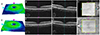 | Figure 4Cirrus HD-OCT of the both eyes two weeks after the first visit. (A-D) Macular Cube 512 × 128 volume scan on Cirrus HD-OCT of the right eye. (E-H) Macular Cube 512 × 128 volume scan on Cirrus HD-OCT of the left eye. No duplication of fovea was noted. No abrupt discontinuation or shadow was noted.
|
References
1. Kim US. Optical coherence tomography. Clin Neuroophthalmol. 2014; 4:17–21.
2. Han IC, Jaffe GJ. Evaluation of artifacts associated with macular spectral-domain optical coherence tomography. Ophthalmology. 2010; 117:1177–1189.e4.
3. Ho J, Sull AC, Vuong LN, et al. Assessment of artifacts and reproducibility across spectral- and time-domain optical coherence tomography devices. Ophthalmology. 2009; 116:1960–1970.
4. Ray R, Stinnett SS, Jaffe GJ. Evaluation of image artifact produced by optical coherence tomography of retinal pathology. Am J Ophthalmol. 2005; 139:18–29.
5. Baskin DE, Gault JA, Vander JF, Dugan JD Jr. Double fovea artifact. Ophthalmology. 2011; 118:429.e1.
6. Kalliath J, Shukla D. Foveal duplication artifact with spectral-domain optical coherence tomography. Ophthalmic Surg Lasers Imaging Retina. 2013; 44:94–96.
7. Behera UC, Shukla D, Kim R. Pseudoduplication of fovea in a human eye. Arch Ophthalmol. 2007; 125:1428–1430.
 XML Download