

 PDF
PDF ePub
ePub Citation
 Print
Print


Abstract
Purpose
To report a case of cytomegalovirus (CMV) corneal endotheliitis following penetrating keratoplasty.
Case summary
A 45-year-old male with a history of re-penetrating keratoplasty due to corneal opacity and graft failure after previous penetrating keratoplasty of his right eye in April 2014, visited our clinic for intermittent injection of the right eye for several weeks (7 months postoperative). Corneal edema, diffuse keratic pigmentation and anterior chamber reaction with decreased endothelial cell density were observed in his right eye using the slit lamp examination. Seven months after keratoplasty, corneal graft rejection were determined but clinical findings showed features of CMV-related corneal endotheliitis. Under the impression of CMV corneal endotheliitis, diagnostic paracentesis was performed for CMV real time polymerase chain reaction (RT-PCR). Additionally, the patient was admitted for intravenous ganciclovir and topical ganciclovir therapy. The next day, the RT-PCR results confirmed CMV infection. After 2 weeks of intravenous ganciclovir treatment, the patient was discharged and prescribed oral ganciclovir for 1 month. A month later, the coin-shaped corneal lesion nearly disappeared. There was no evidence of complication or recurrence.
Conclusions
CMV corneal endotheliitis typically presents with coin-shaped keratic pigmentation and can be confirmed with RT-PCR using aqueous humor collected from the anterior chamber. Due to the long period of systemic and topical steroid therapy, the risk of viral endotheliitis is relatively high in patients with a history of penetrating keratoplasty. Corneal graft rejection is similar to corneal endotheliitis in symptoms and clinical features such as ciliary injection, decreased visual acuity, corneal edema or anterior chamber reaction. In patients after penetrating keratoplasty, CMV RT-PCR should be considered if the clinical features suggest viral endotheliitis.
References
1. Koizumi N, Suzuki T, Uno T, et al. Cytomegalovirus as an etiologic factor in corneal endotheliitis. Ophthalmology. 2008; 115:292–7.e3.

2. Sonoyama H, Araki-Sasaki K, Osakabe Y, et al. Detection of abdominal DNA from cytomegalovirus corneal endotheliitis abdominal penetrating keratoplasty. Cornea. 2010; 29:683–5.
3. Taylor-Wiedeman J, Sissons JG, Borysiewicz LK, Sinclair JH. Monocytes are a major site of persistence of human abdominal in peripheral blood mononuclear cells. J Gen Virol. 1991; 72(Pt 9):2059–64.
4. Slobedman B, Stern JL, Cunningham AL, et al. Impact of human cytomegalovirus latent infection on myeloid progenitor cell gene expression. J Virol. 2004; 78:4054–62.
5. Nakamura T, Ishikawa F, Sonoda KH, et al. Characterization and distribution of bone marrow-derived cells in mouse cornea. Invest Ophthalmol Vis Sci. 2005; 46:497–503.
6. Holland EJ, Bennett SR, Brannian R, et al. The risk of abdominal transmission by penetrating keratoplasty. Am J Ophthalmol. 1988; 105:357–60.
7. Suzuki T, Ohashi Y. Corneal endotheliitis. Semin Ophthalmol. 2008; 23:235–40.
8. Kandori M, Inoue T, Takamatsu F, et al. Prevalence and features of keratitis with quantitative polymerase chain reaction positive for cytomegalovirus. Ophthalmology. 2010; 117:216–22.
9. Yutthitham K, Ruamviboonsuk P. The high-dose, alternate-week intravitreal ganciclovir injections for cytomegalovirus retinitis in acquired immune deficiency syndrome patients on highly active antiretroviral therapy. J Med Assoc Thai. 2005; 88(Suppl 9):S63–8.
10. Ausayakhun S, Yuvaves P, Ngamtiphakom S, Prasitsilp J. Treatment of cytomegalovirus retinitis in AIDS patients with abdominal ganciclovir. J Med Assoc Thai. 2005; 88(Suppl 9):S15–20.
11. Arevalo JF, Garcia RA, Mendoza AJ. High-dose (5000-microg) abdominal ganciclovir combined with highly active antiretroviral therapy for cytomegalovirus retinitis in HIV-infected patients in Venezuela. Eur J Ophthalmol. 2005; 15:610–8.
12. Su CC, Wang IJ, Chen WL, et al. Topical ganciclovir treatment in patients with cytomegalovirus endotheliitis receiving penetrating keratoplasty. Clin Experiment Ophthalmol. 2013; 41:339–47.
13. Choi WS, Cho JH, Kim HK, et al. A case of CMV endotheliitis treated with intravitreal ganciclovir injection. Korean J Ophthalmol. 2013; 27:130–2.
14. Panda A, Vanathi M, Kumar A, et al. Corneal graft rejection. Surv Ophthalmol. 2007; 52:375–96.
15. Kim EC, Kim MS. Graft rejection in penetrating keratoplasty. J Korean Ophthalmol Soc. 2003; 44:289–95.
16. Koizumi N, Inatomi T, Suzuki T, et al. Clinical features and abdominal of cytomegalovirus corneal endotheliitis: analysis of 106 cases from the Japan corneal endotheliitis study. Br J Ophthalmol. 2015; 99:54–8.
17. Chee SP, Jap A, Ling EC, Ti SE. Cytomegalovirus-positive corneal stromal edema with keratic precipitates after penetrating abdominal: a case-control study. Cornea. 2013; 32:1094–8.
18. Chee SP, Bacsal K, Jap A, et al. Clinical features of abdominal anterior uveitis in immunocompetent patients. Am J abdominal. 2008; 145:834–40.
19. Koizumi N, Yamasaki K, Kawasaki S, et al. Cytomegalovirus in aqueous humor from an eye with corneal endotheliitis. Am J Ophthalmol. 2006; 141:564–5.
Figure 1.
Clinical manifestations of cytomegalovirus corneal endotheliitis examined by slit-lamp biomicroscopy. Pre-therapeutic photograph. Diffuse coin-shped keratic precipitates and stromal corneal edema are observed (arrows).
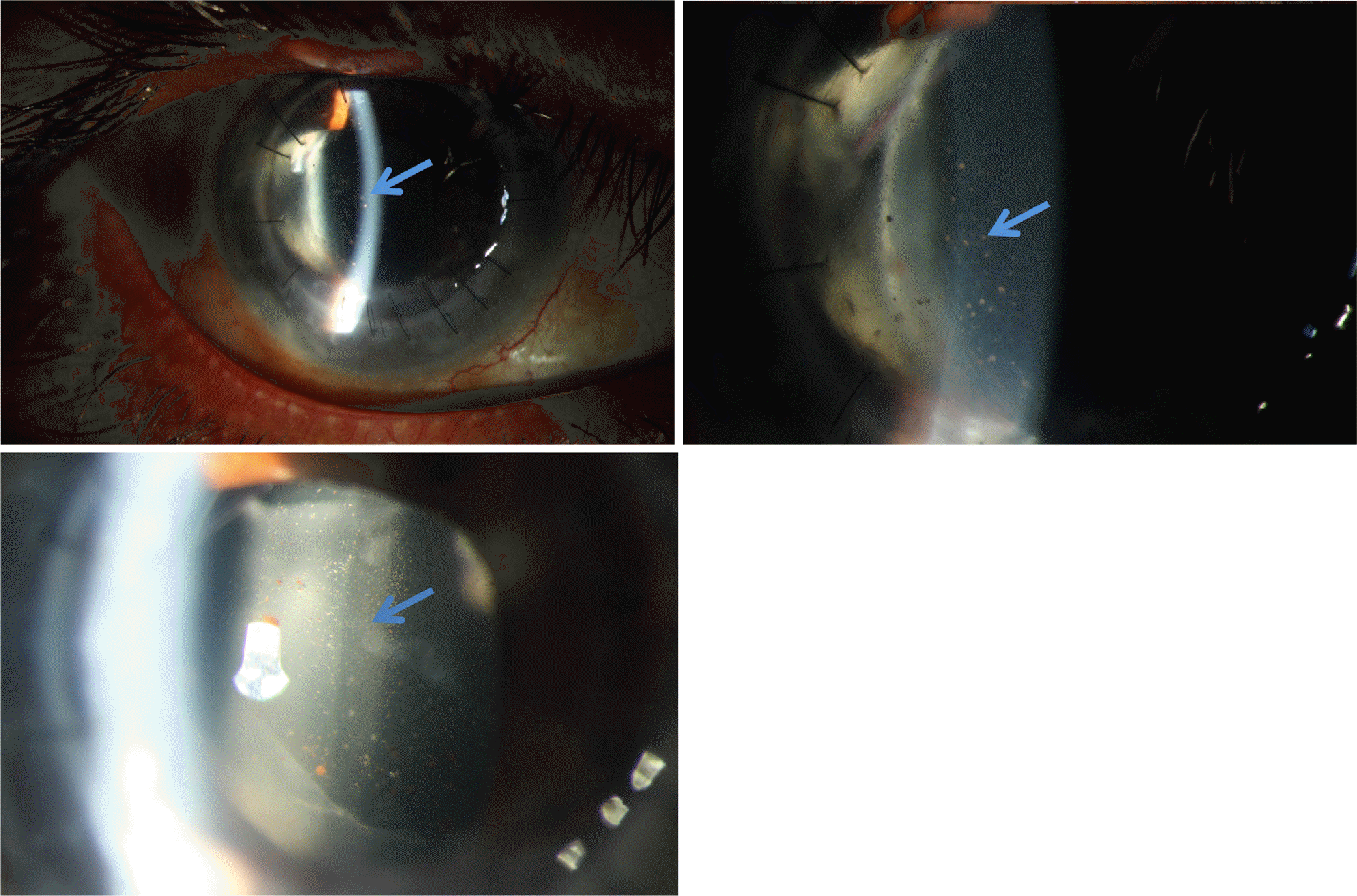
Figure 2.
Clinical finding 7 months after gancyclovir therapy. Coin-shaped keratic pigmentation disappeared and no anterior chamber reaction was remained.
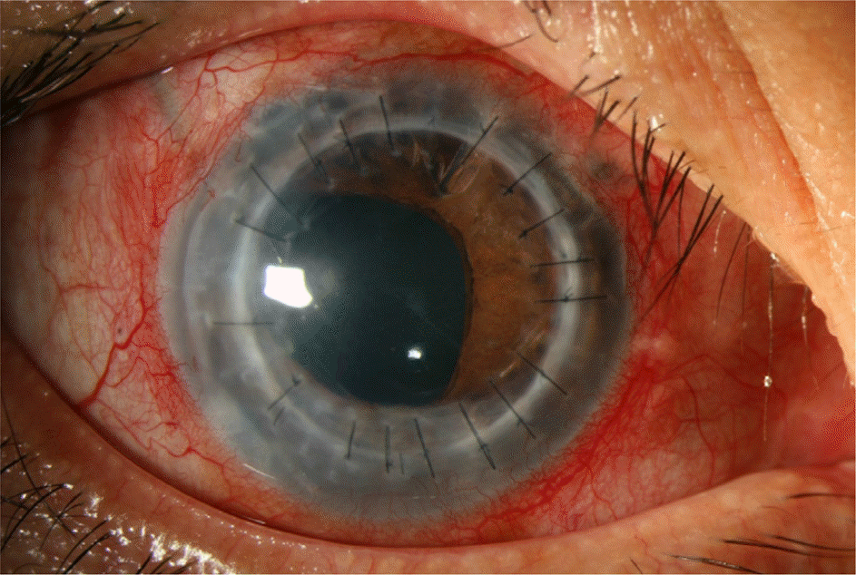
Table 1.
Follow up visual acuity, IOP, specular, CCT
 XML Download