Abstract
Purpose
To report four cases of split cornea transplantation involving separate Descemet membrane keratoplasty and Deep anterior lamella keratoplasty from a single cornea.
Case summary
Four donor corneas were separated into the endothelium and other layers. The endothelial layers were transplanted into 4 pseudophakic bullous keratopathy patients, and the other layers were stored in corneal storage media. Deep anterior lamella keratoplasties were performed with the stored corneas in 2 lipid keratopathy and 2 keratoconus patients. Postoperatively, all grafted corneas were stable.
Conclusions
The authors report the first 4 cases of split cornea transplantation in Korea, which is experiencing a shortage of do-nated corneas. Split cornea transplantation will be of benefit to a large number of patients by separating a single cornea into separate layers to be implanted into two patients.
References
1. Archila EA. Deep lamellar keratoplasty dissection of host tissue with intrastromal air injection. Cornea. 1984–1985; 3:217–8.

2. Shimazaki J, Shimmura S, Ishioka M, Tsubota K. Randomized clinical trial of deep lamellar keratoplasty vs penetrating keratoplasty. Am J Ophthalmol. 2002; 134:159–65.
3. Tsubota K, Kaido M, Monden Y, et al. A new surgical technique for deep lamellar keratoplasty with single running suture adjustment. Am J Ophthalmol. 1998; 126:1–8.

4. Melles GR, Ong TS, Ververs B, van der Wees J. Descemet abdominal endothelial keratoplasty (DMEK). Cornea. 2006; 25:987–90.
5. Ang M, Wilkins MR, Mehta JS, Tan D. Descemet membrane abdominal keratoplasty. Br J Ophthalmol. 2015; May:pii: bjoph-thalmol-2015–306837. [Epub ahead of print].
6. Güell JL, Morral M, Gris O, et al. Bimanual technique for insertion and positioning of endothelium-Descemet membrane graft in Descemet membrane endothelial keratoplasty. Cornea. 2013; 32:1521–6.

7. Satué M, Rodríguez-Calvo-de-Mora M, Naveiras M, et al. Standardization of the Descemet membrane endothelial keratoplasty technique: Outcomes of the first 450 consecutive cases. Arch Soc Esp Oftalmol. 2015; 90:356–64.

8. Terry MA, Straiko MD, Veldman PB, et al. Standardized DMEK technique: reducing complications using prestripped tissue, novel glass injector, and sulfur hexafluoride (SF6) gas. Cornea. 2015; 34:845–52.
9. Weller JM, Tourtas T, Kruse FE, et al. Descemet membrane abdominal keratoplasty as treatment for graft failure after descemet stripping automated endothelial keratoplasty. Am J Ophthalmol. 2015; 159:1050–7.e2.
10. Korean Network for Organ Sharing (KONOS). 2014 Annual Data Report. Seoul: KONOS;2014. 11:p. 135–71.
11. Department of Ophthalmology and Visual Science, College of Medicine, The Catholic University of Korea. 2014 Annual Report. Seoul: The Catholic University of Korea;2014. p. 140–146.
12. Vajpayee RB, Sharma N, Jhanji V, et al. One donor cornea for 3 abdominal: a new concept for corneal transplantation surgery. Arch Ophthalmol. 2007; 125:552–4.
13. Kim ST, Lee YC, Heo J, et al. Various treatments using invaluable donor cornea. J Korean Ophthalmol Soc. 2009; 50:471–6.

14. Ple-Plakon PA, Shtein RM. Trends in corneal transplantation: abdominal and techniques. Curr Opin Ophthalmol. 2014; 25:300–5.
15. Kymionis GD, Mikropoulos DG, Portaliou DM, et al. New abdominal on lamellar keratoplasty. Adv Ther. 2014; 31:494–511.
16. Tausif HN, Johnson L, Titus M, et al. Corneal donor tissue abdominal for Descemet's membrane endothelial keratoplasty. J Vis Exp. 2014; 91:51919.
17. Kruse FE, Laaser K, Cursiefen C, et al. A stepwise approach to abdominal preparation and insertion increases safety and outcome of Descemet membrane endothelial keratoplasty. Cornea. 2011; 30:580–7.
18. Lie JT, Birbal R, Ham L, et al. Donor tissue preparation for Descemet membrane endothelial keratoplasty. J Cataract Refract Surg. 2008; 34:1578–83.

19. Lee K, Boimer C, Hershenfeld S, et al. Sustainability of Routine Notification and Request legislation on eye bank tissue supply and corneal transplantation wait times in Canada. Can J Ophthalmol. 2011; 46:381–5.

20. Ang M, Lim F, Htoon HM, et al. Visual acuity and contrast abdominal following Descemet stripping automated endothelial abdominal. Br J Ophthalmol. 2015; Jul:pii: bjophthalmol-2015–306975. [Epub ahead of print].
21. Yum HR, Kim MS, Kim EC. Retrocorneal membrane after abdominal membrane endothelial keratoplasty. Cornea. 2013; 32:1288–90.
22. Park CY, Chuck RS. Non-Descemet stripping Descemet membrane endothelial keratoplasty. Cornea. 2013; 32:1607–9.

23. Heindl LM, Riss S, Bachmann BO, et al. Split cornea abdominal for 2 recipients: a new strategy to reduce corneal tissue cost and shortage. Ophthalmology. 2011; 118:294–301.
24. Heindl LM, Riss S, Laaser K, et al. Split cornea transplantation for 2 recipients – review of the first 100 consecutive patients. Am J Ophthalmol. 2011; 152:523–32.e2.

25. Heindl LM, Cursiefen C. Split-cornea transplantation – a novel concept to reduce corneal donor shortage. Klin Monbl Augenheilkd. 2012; 229:608–14.
26. Heindl LM, Riss S, Adler W, et al. Split cornea transplantation: abdominalship between storage time of split donor tissue and outcome. Ophthalmology. 2013; 120:899–907.
27. Melles GR, Ong TS, Ververs B, van der Wees J. Preliminary abdominal results of Descemet membrane endothelial keratoplasty. Am J Ophthalmol. 2008; 145:222–7.
28. Ham L, Dapena I, van Luijk C, et al. Descemet membrane abdominal keratoplasty (DMEK) for Fuchs endothelial dystrophy: review of the first 50 consecutive cases. Eye (Lond). 2009; 23:1990–8.
29. Ham L, van Luijk C, Dapena I, et al. Endothelial cell density after Descemet membrane endothelial keratoplasty: 1- to 2-year fol-low-up. Am J Ophthalmol. 2009; 148:521–7.

30. Price MO, Giebel AW, Fairchild KM, Price FW Jr. Descemet's membrane endothelial keratoplasty: prospective multicenter study of visual and refractive outcomes and endothelial survival. Ophthalmology. 2009; 116:2361–8.
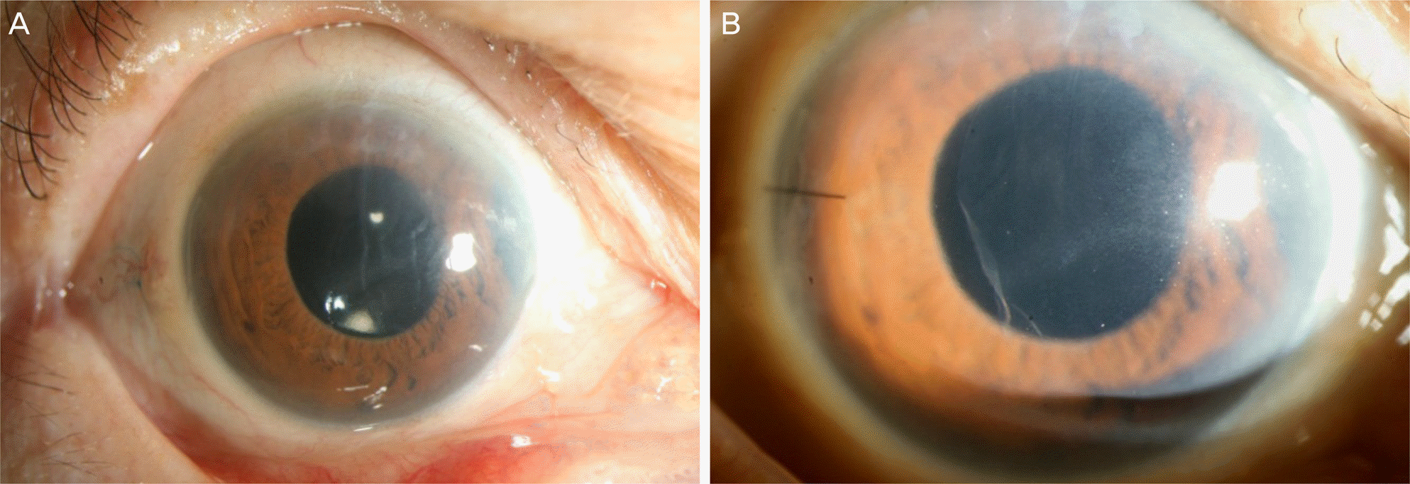
Figure 1.
Slit-lamp photographs of a Descemet membrane keratoplasty (DMEK) case. (A) Corneal opacity is seen in pseudophakic bullous keratopathy (PBK) patient before DMEK by slit lamp examination. (B) Clear graft is seen after two months following DMEK.

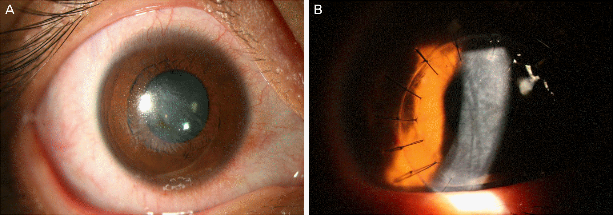
Figure 2.
Slit-lamp photographs of a Deep anterior lamella keratoplasty (DALK) case. (A) Corneal opacity is seen at the center of the cornea in the keratoconus patient before DALK by slit lamp examination. (B) After one month postoperatively, clear graft but mild descemet membrane folds are to be seen in the recipient cornea. There is no rejection sign.

Table 1.
Demographic and operative details of DMEK patients
Table 2.
Demographic and operative details of DALK patients


 PDF
PDF ePub
ePub Citation
 Print
Print


 XML Download