

 PDF
PDF ePub
ePub Citation
 Print
Print


Abstract
Methods
We performed a retrospective chart review analysis of patients referred to the neuro-ophthalmology clinic due to unknown etiology of decreased visual acuity and diagnosed with oil droplet cataract. Clinical features including history, result of ophthalmologic examinations, and clinical course were evaluated.
Results
Among the patients referred to the neuro-ophthalmology clinic due to unknown etiology of decreased visual acuity, 6 patients were diagnosed with oil droplet cataract. The patients ranged from 38 to 63 years of age and their best corrected visual acuities at their first visits were between 0.1 and 0.7. Ophthalmologic examinations including neuro-ophthalmologic tests were normal except for changes in lens nucleus and peculiar fundus reflexes were observed using retinoscopy in all patients. Five eyes of 4 patients underwent cataract surgery and all 5 eyes achieved the best corrected visual acuity of 1.0 or higher.
Conclusions
Oil droplet cataract is a cause of decreased visual acuity of unknown etiology that can be missed. The disease abnormalities are difficult to observe because only subtle changes in lens nucleus are apparent on slit lamp examination; however characteristic fundus reflexes can be identified using retinoscopy. Ophthalmologists should thoroughly understand the oil droplet cataract and diagnose it in the early stages to avoid misdiagnosis and unnecessary costs.
Go to : 
References
1. Eifrig DE. Identifying oil-drop cataracts. J Cataract Refract Surg. 1994; 20:105–6.

2. Soong HK, Dastjerdi MH. Lenticular myopia from oil-drop abdominal: a cautionary note in laser in situ keratomileusis. J Cataract Refract Surg. 2004; 30:2438–40.
3. Mohammadpour M, Jabbarvand M. Oil-drop cataracts. J Cataract Refract Surg. 2008; 34:1425.
4. Heher KL, Stark WJ, Miller NR. Oil-drop cataracts. J Cataract Refract Surg. 1993; 19:306–8.
5. Rubin ML. The case of the myopic capitalist. Surv Ophthalmol. 1968; 13:122–3.
6. Lee KE, Klein BE, Klein R, Wong TY. Changes in refraction over 10 years in an adult population: the Beaver Dam Eye study. Invest Ophthalmol Vis Sci. 2002; 43:2566–71.
7. Wu Z, Lim JI, Sadda SR. Axial length: a risk factor for cataractogenesis. Ann Acad Med Singapore. 2006; 35:416–9.
8. Wong TY, Foster PJ, Johnson GJ, Seah SK. Refractive errors, axial ocular dimensions, and age-related cataracts: the Tanjong Pagar survey. Invest Ophthalmol Vis Sci. 2003; 44:1479–85.
9. Guzowski M, Wang JJ, Rochtchina E, et al. Five-year refractive changes in an older population: the Blue Mountains Eye Study. Ophthalmology. 2003; 110:1364–70.
10. Cho YK, Huang W, Nishimura E. Myopic refractive shift represents dense nuclear sclerosis and thin lens in lenticular myopia. Clin Exp Optom. 2013; 96:479–85.
11. Samarawickrama C, Wang JJ, Burlutsky G, et al. Nuclear cataract and myopic shift in refraction. Am J Ophthalmol. 2007; 144:457–9.
12. Brown NP, Koretz JF, Bron AJ. The development and maintenance of emmetropia. Eye (Lond). 1999; 13(Pt 1):83–92.
13. Brown NA, Hill AR. Cataract: the relation between myopia and cataract morphology. Br J Ophthalmol. 1987; 71:405–14.
14. Kaufman BJ, Sugar J. Discrete nuclear sclerosis in young patients with myopia. Arch Ophthalmol. 1996; 114:1178–80.
15. Kuroda T, Fujikado T, Maeda N, et al. Wavefront analysis in eyes with nuclear or cortical cataract. Am J Ophthalmol. 2002; 134:1–9.
16. Congdon NG, Taylor HR. Age-related cataract. Johnson GJ, Minassian DC, West S, Weale R, editors. The Epidemiology of Eye Disease. 2nd ed.London: London;2003. chap. 8.
17. Kashima K, Trus BL, Unser M, et al. Aging studies on normal lens using the Scheimpflug abdominal camera. Invest Ophthalmol Vis Sci. 1993; 34:263–9.
18. Panchapakesan J, Rochtchina E, Mitchell P. Myopic refractive shift caused by incident cataract: the Blue Mountains Eye Study. Ophthalmic Epidemiol. 2003; 10:241–7.
19. van Heyningen R. What happens to the human lens in cataract. Sci Am. 1975; 233:70–2. 77–81.
Go to :
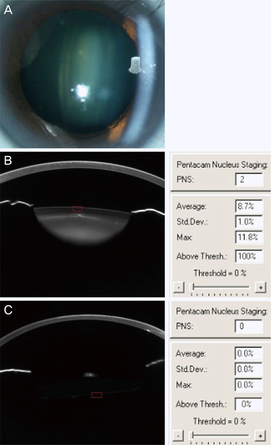 | Figure 1.Comparison of lens status between patients and normal controls. Slit lamp photographs (A) and scheimpflug image of the right eye in patient 1 (B). The center of nucleus is relatively homogeneously foggy compared to the fairly clear cortex. The average and maximal opacity of the lens were 8.7% and 11.8%. Pentacam nucleus staging was stage 2. In normal eye, the average and maximal opacity of lens were 0% (C). PNS =Pentacam Nucleus Staging; Std. Dev = standard deviation. |
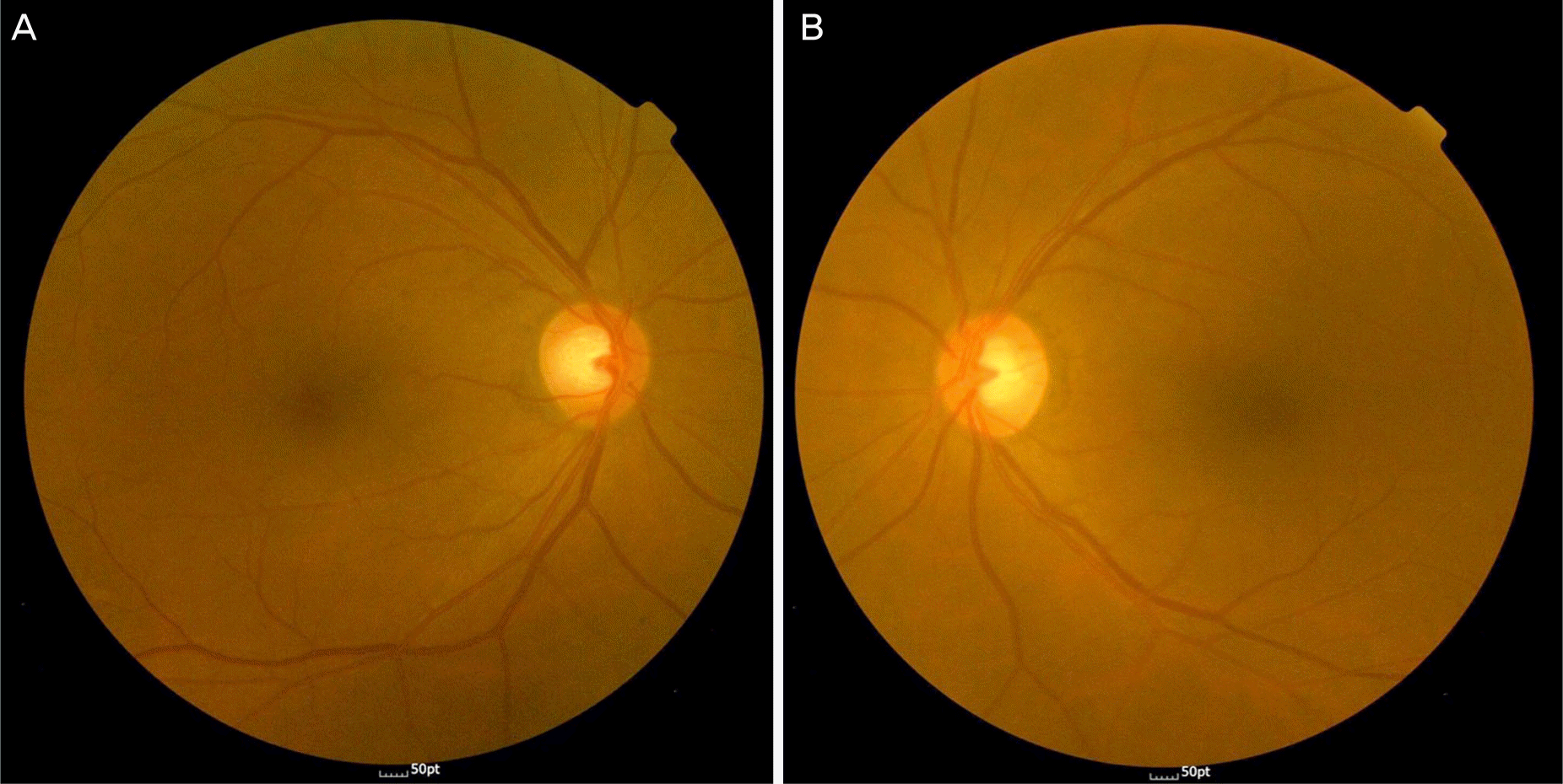 | Figure 2.Fundus photography of patient 2. The left eye has oil droplet cataract. But the right eye has a clear lens. The view of the left fundus (B) is not hazy compared to the right fundus (A). |
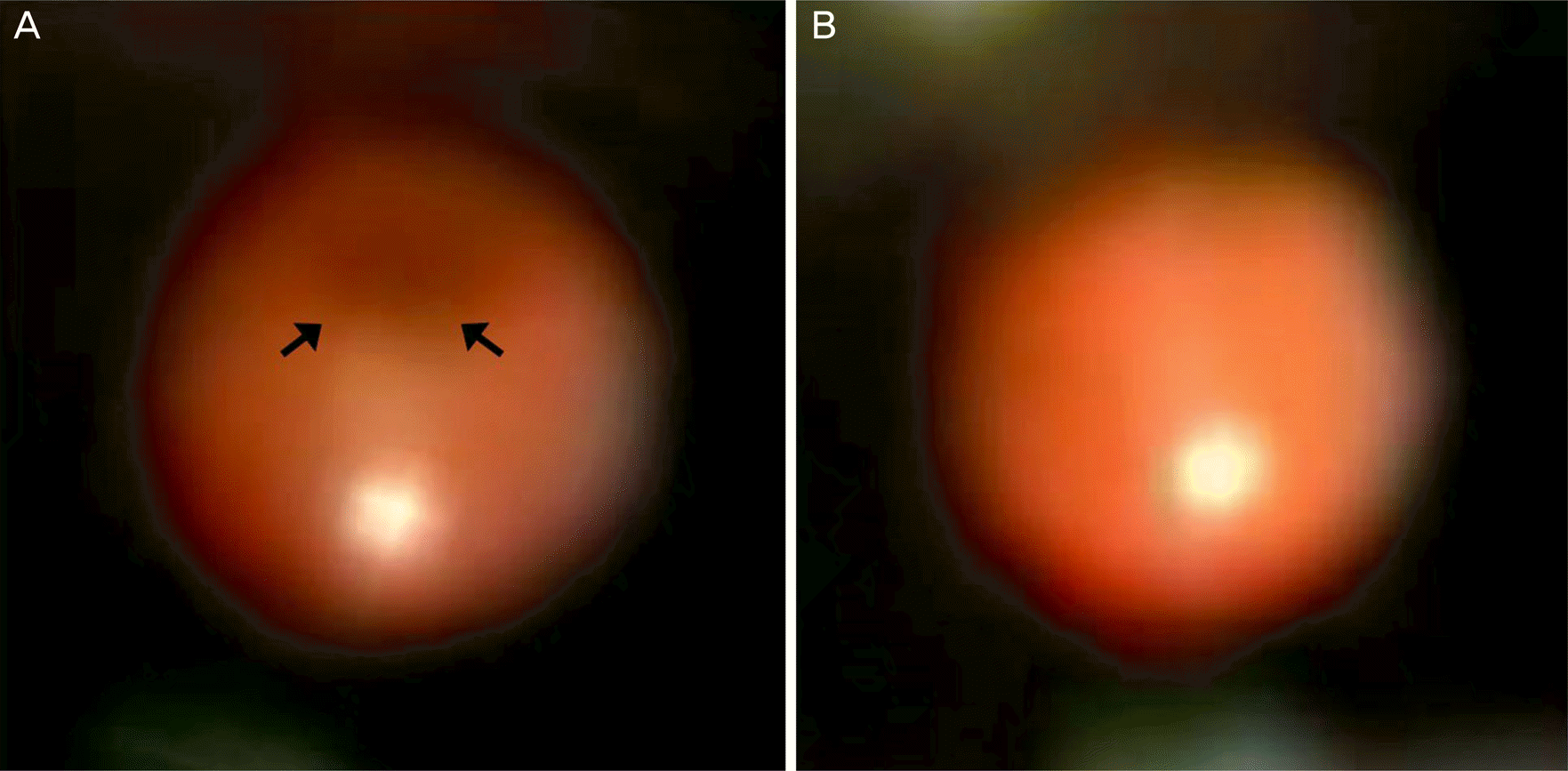 | Figure 3.Comparison of fundus reflex between patients and normal controls. Retinoscopy can identify characteristic dark silluette (arrows) fundus reflexes (A) in contrast with normal reflex (B). |
Table 1.
Demographics of patients
| Patient | Age (years) | Gender | Laterality | Disease status | Initial UCVA | Initial BCVA | Axial length (mm) | Autorefraction (diopters, SE) |
|---|---|---|---|---|---|---|---|---|
| 1* | 38 | Female | OD | Oil droplet cataract | 0.2 | 0.7 | 25.01 | −14.5 |
| OS | Oil droplet cataract | 0.05 | 0.2 | 25.49 | −13.0 | |||
| 2* | 55 | Female | OS | Oil droplet cataract | 0.4 | 0.5 | 23.01 | −4.0 |
| OD | Normal | 1.2 | 1.2 | 23.20 | 0.25 | |||
| 3* | 63 | Male | OD | Oil droplet cataract | 0.02 | 0.4 | 24.82 | −6.0 |
| OS | Normal | 0.4 | 1.0 | 24.67 | −1.5 | |||
| 4 | 51 | Female | OD | Oil droplet cataract | 0.3 | 0.4 | – | −0.5 |
| OS | Normal | 0.6 | 0.8 | – | 0.25 | |||
| 5 | 62 | Female | OD | Oil droplet cataract | 0.02 | 0.1 | – | −9.0 |
| OS | Normal | 0.2 | 0.4 | – | −5.0 | |||
| 6* | 50 | Male | OD | Oil droplet cataract | 0.04 | 0.5 | 28.53 | −12.0 |
| OS | Normal | 0.04 | 1.0 | 29.04 | −6.0 |
Table 2.
Changes of visual acuity after phacoemulsification and posterior chamber intraocular lens implantation
 XML Download