

 PDF
PDF ePub
ePub Citation
 Print
Print


Abstract
Purpose
To evaluate the clinical effectiveness of pneumatic retinopexy as a treatment method for pseudophakic retinal detachment.
Methods
A retrospective chart review was conducted of medical records of 38 patients who underwent pneumatic retinopexy using SF6 gas from January 2003 to December 2011 and who were observed during a follow-up period longer than 6 months. Primary and final success rates and final visual acuity were analyzed. Primary success was defined as retinal attachment at the last visit without additional surgery. Final success was defined as retinal reattachment at the last visit regardless of additional surgery.
Results
The mean patient age was 58.47 ± 17.00 years. All retinal tears were located in the upper retina (from 8 to 4 o'clock). Preoperative mean visual acuity was 1.17 ± 1.00 log MAR, and postoperative mean visual acuity was 0.42 ± 0.48 log MAR. The primary success rate was 61%, and patients with re-detached retina underwent repeat pneumatic retinopexy or other surgery such as scleral buckling or pars plana vitrectomy. At the final visit, all of the patients demonstrated successful results.
Go to : 
References
1. The Retina Society Terminology Committee. The classification of retinal detachment with proliferative vitreoretinopathy. abdominalogy. 1983; 90:121–5.
2. Schepens CL. Retinal detachment and aphakia. AMA Arch abdominal. 1951; 45:1–17.

3. Norton EW. Retinal detachment in aphakia. Trans Am Ophthalmol Soc. 1963; 61:770–89.
4. Haimann MH, Burton TC, Brown CK. Epidemiology of retinal detachment. Arch Ophthalmol. 1982; 100:289–92.
5. Yoshida A, Ogasawara H, Jalkh AE, et al. Retinal detachment after cataract surgery. Predisposing factors. Ophthalmology. 1992; 99:453–9.
6. Yoshida A, Ogasawara H, Jalkh AE, et al. Retinal detachment after cataract surgery. Surgical results. Ophthalmology. 1992; 99:460–5.
7. Freeman HM, Dobbie JG, Friedman MW. Pseudophakic retinal detachment. Mal Probl Ophthalmol. 1979; 20:345–53.
8. Javitt JC, Street DA, Tielsch JM, et al. National outcomes of abdominal extraction. Retinal detachment and endophthalmitis after outpatient cataract surgery. Cataract Patient Outcomes Research Team. Ophthalmology. 1994; 101:100–5. discussion 106.
9. Ninn-Pedersen K, Bauer B. Cataract patients in a defined Swedish population, 1986 to 1990. V. Postoperative retinal detachments. Arch Ophthalmol. 1996; 114:382–6.
10. Wegener M, Alsbirk PH, Højgaard-Olsen K. Outcome of 1000 consecutive clinic- and hospital-based cataract surgeries in a Danish county. J Cataract Refract Surg. 1998; 24:1152–60.
11. Olsen G, Olson RJ. Update on a long-term, prospective study of capsulotomy and retinal detachment rates after cataract surgery. J Cataract Refract Surg. 2000; 26:1017–21.
12. Bobrow JC. Visual outcomes after anterior vitrectomy: abdominal of ECCE and phacoemulsification. Trans Am Ophthalmol Soc. 1999; 97:281.
13. Yap EY, Heng WJ. Visual outcome and complications after abdominal capsule rupture during phacoemulsification surgery. Int abdominal. 1999; 23:57–60.
14. Ho PC, Tolentino FI. Pseudophakic retinal detachment. Surgical abdominal rate with various types of IOLs. Ophthalmology. 1984; 91:847–52.
15. Wilkinson CP. Pseudophakic retinal detachments. Retina. 1986; 5:1–4.
16. Han DP, Mohsin NC, Guse CE, et al. Comparison of pneumatic abdominal and scleral buckling in the management of primary abdominal retinal detachment. Southern Wisconsin Pneumatic Retinopexy Study Group. Am J Ophthalmol. 1998; 126:658–68.
17. Chen JC, Robertson JE, Coonan P, et al. Results and complications of pneumatic retinopexy. Ophthalmology. 1988; 95:601–6.
18. Tornambe PE, Hilton GF. Pneumatic retinopexy. A multicenter randomized controlled clinical trial comparing pneumatic abdominal with scleral buckling. The Retinal Detachment Study Group. Ophthalmology. 1989; 96:772–83. discussion 784.
19. Lee SE, Chang MH. The success rate and factors influencing the results of pneumatic retinopexy. J Korean Ophthalmol Soc. 2013; 54:1241–7.
20. Hilton GF, Grizzard WS. Pneumatic retinopexy. A two-step outpatient operation without conjunctival incision. Ophthalmology. 1986; 93:626–41.
21. Poliner LS, Grand MG. Clinical experience. Torname PE, Grizzard SW, Vygantas CM, editors. Pneumatic Retinopexy: A Clinical Symposium. Des Plaines: Greenwood Publishing;1989. chap. 3.
22. Dominguez DA, Boyd BF, Gordon S. Repeated insufflation of ex-pansive gas. Highlights Ophthalmol Lett. 1986; 14:1–14.
23. Cho WH, Lee DC, Chang MH. Three cases of pneumoretinopexy for rhegmatogenous retinal detachment by multiple retinal tears over 1 hour in distance. J Korean Ophthalmol Soc. 2005; 46:2110–4.
24. Brinton DA, Hilton GF. Pneumatic retinopexy and alternative abdominall detachment techniques. Ryan SJ, Wilkinson CP, editors. Retina. 3rd ed.St. Louis: St. Louis;2001. 3:chap. 130.
25. Hilton GF, Das T, Majji AB, Jalali S. Pneumatic retinopexy: abdominal and practice. Indian J Ophthalmol. 1996; 44:131–43.
26. Kleinmann G, Rechtman E, Pollack A, et al. Pneumatic retinopexy: results in eyes with classic vs relative indications. Arch abdominal. 2002; 120:1455–9.
27. Bartz-Schmidt KU, Kirchhof B, Heimann K. Primary vitrectomy for pseudophakic retinal detachment. Br J Ophthalmol. 1996; 80:346–9.
28. Brazitikos PD, Androudi S, Christen WG, Stangos NT. Primary pars plana vitrectomy versus scleral buckle surgery for the abdominal of pseudophakic retinal detachment: a randomized clinical trial. Retina. 2005; 25:957–64.
29. Brazitikos PD, D'Amico DJ, Tsinopoulos IT, Stangos NT. Primary vitrectomy with perfluoro-n-octane use in the treatment of abdominal retinal detachment with undetected retinal breaks. Retina. 1999; 19:103–9.
30. Campo RV, Sipperley JO, Sneed SR, et al. Pars plana vitrectomy without scleral buckle for pseudophakic retinal detachments. Ophthalmology. 1999; 106:1811–5. discussion 1816.
31. Weichel ED, Martidis A, Fineman MS, et al. Pars plana vitrectomy versus combined pars plana vitrectomy-scleral buckle for primary repair of pseudophakic retinal detachment. Ophthalmology. 2006; 113:2033–40.
32. Speicher MA, Fu AD, Martin JP, von Fricken MA. Primary abdominal alone for repair of retinal detachments following cataract surgery. Retina. 2000; 20:459–64.
33. Martínez-Castillo V, Boixadera A, Verdugo A, García-Arumi J. Pars plana vitrectomy alone for the management of inferior breaks in pseudophakic retinal detachment without facedown position. Ophthalmology. 2005; 112:1222–6.
34. Heimann H, Zou X, Jandeck C, et al. Primary vitrectomy for abdominal retinal detachment: an analysis of 512 cases. Graefes Arch Clin Exp Ophthalmol. 2006; 244:69–78.
35. Devenyi RG, de Carvalho Nakamura H. Combined scleral buckle and pars plana vitrectomy as a primary procedure for pseudophakic retinal detachments. Ophthalmic Surg Lasers. 1999; 30:615–8.
36. Brazitikos PD. The expanding role of primary pars plana abdominal in the treatment of rhegmatogenous noncomplicated abdominall detachment. Semin Ophthalmol. 2000; 15:65–77.
Go to :
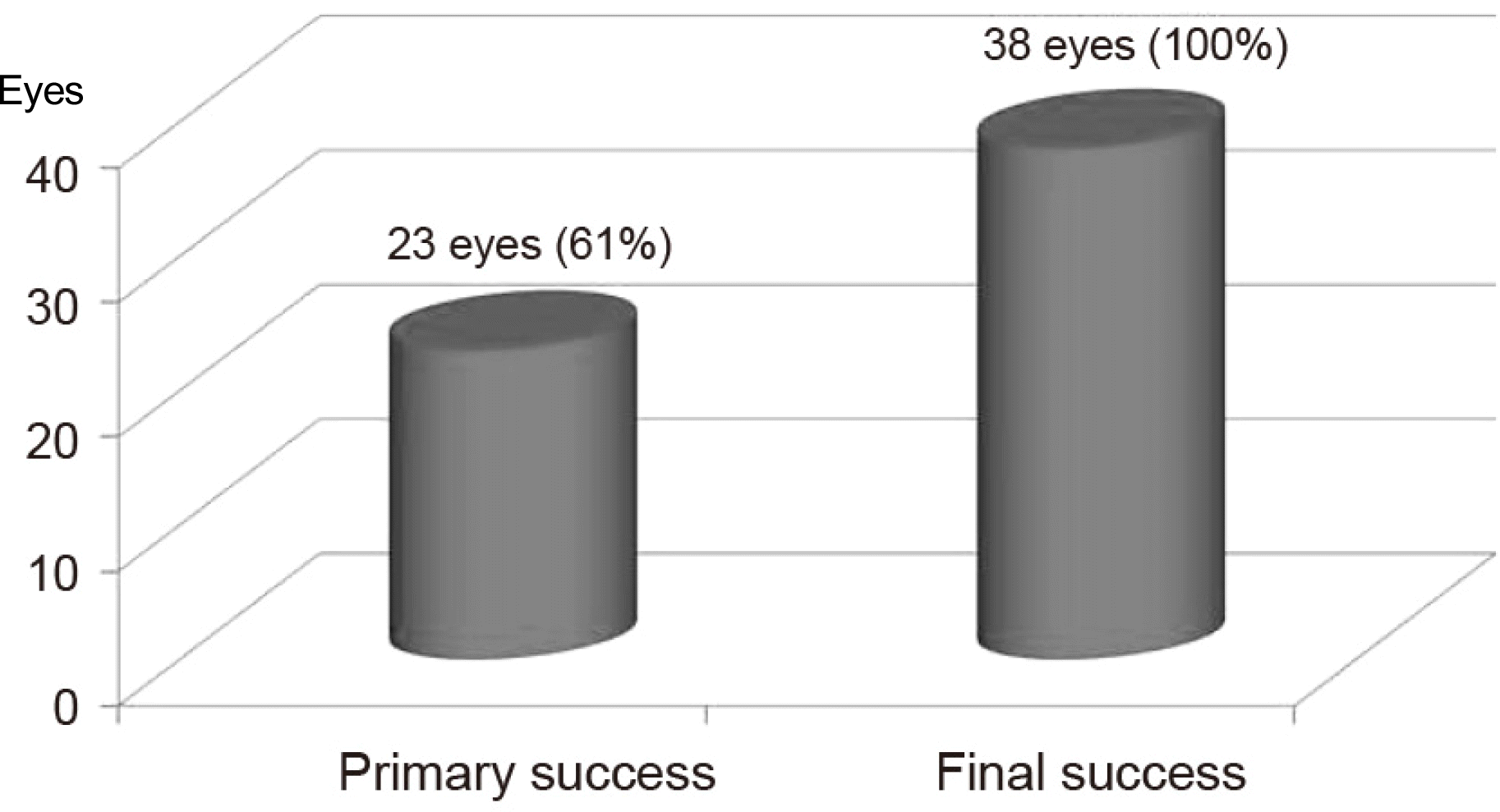 | Figure 1.Success rate of pneumatic retinopexy. The primary success rate was 61% and the final success rate was 100% with additional surgery as scleral buckling or pars plana vitrectomy in redetachment patients. |
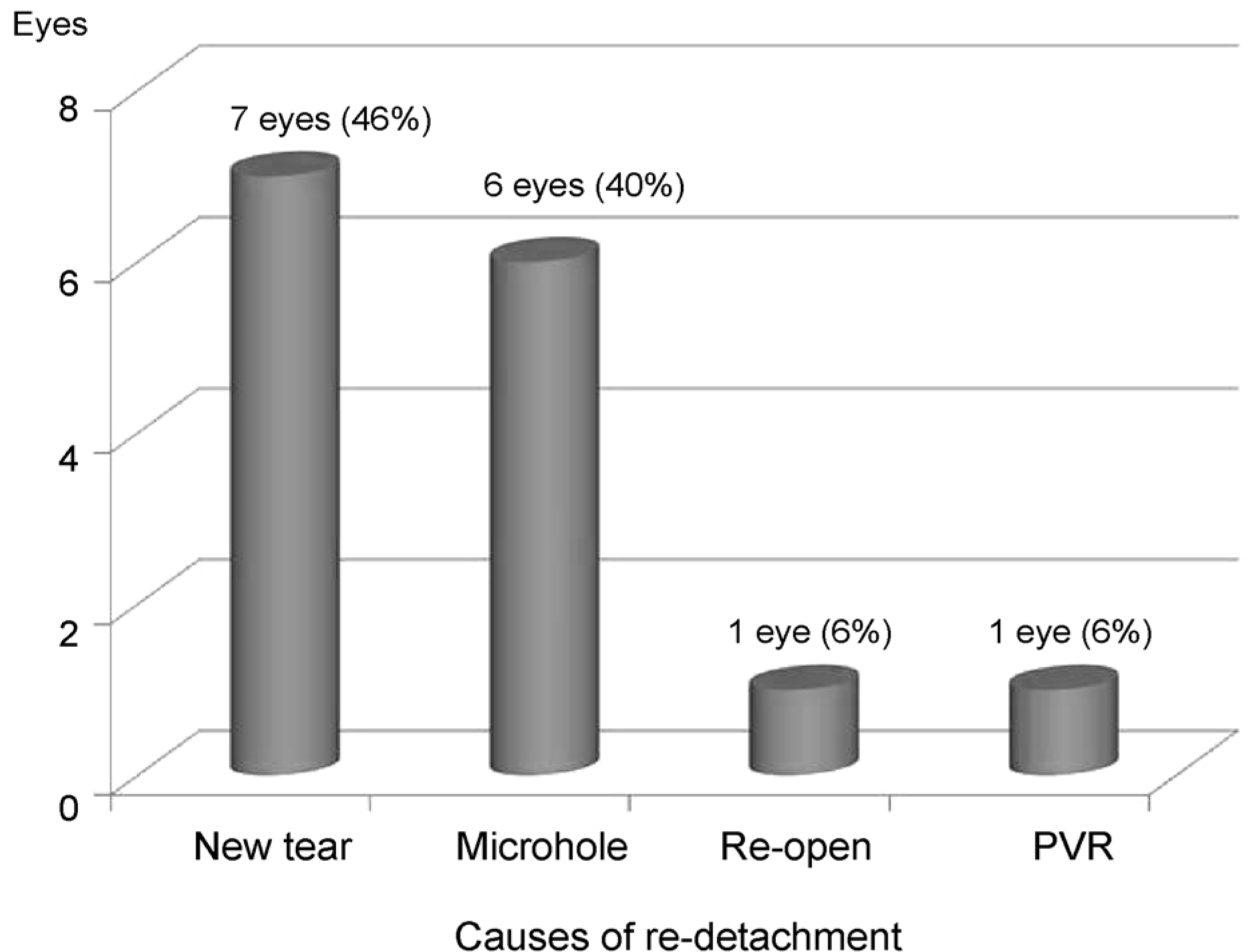 | Figure 2.Causes of redetachment. Causes of Re-detachment after the pneumatic retinopexy as a primary treatment for pseudophakic retinal detachment were new tear, microhole, re-opening tear, proliferative vitreoretinopathy (PVR). |
Table 1.
Demographic and clinical data
 XML Download