

 PDF
PDF ePub
ePub Citation
 Print
Print


Abstract
Purpose
To evaluate the effect of cataract surgery on subfoveal choroidal thickness (SCT) and investigate the relationship between the variation of SCT and refractive error.
Methods
We retrospectively reviewed the medical records of 47 patients (47 eyes) who underwent uneventful phacoemulsification cataract surgery from March 2012 to February 2014. SCTs were measured using spectraldomain optical coherence tomography performed before surgery and at 1 month, 3 months and 6 months postoperatively. We investigated the differences in target refraction (TR) and postoperative spherical equivalent (SE), intraocular pressure (IOP) and central macular thickness (CMT) at all follow-ups.
Results
Compared with preoperative measurements, SCT showed a significant increase of 5.9 ± 13.3 μ m at postoperative 1 month and 7.6 ± 18.1 μ m at postoperative 3 months (p = 0.004 and p = 0.006, respectively), but no significant differences at post-operative 6 months (p = 0.104). The correlation between the variation of SCT and the differences in postoperative SE and TR were not significant at 1 month and 6 months, but were positively significant at 3 months (r = 0.310, p = 0.034). The variation of SCT showed no significant correlations with the postoperative change in IOP and CMT.
Go to : 
References
1. Grossniklaus HE, Green WR. Choroidal neovascularization. Am J Ophthalmol. 2004; 137:496–503.

2. Iida T, Kishi S, Hagimura N, Shimizu K. Persistent and bilateral choroidal vascular abnormalities in central serous chorioretinopathy. Retina. 1999; 19:508–12.
3. Torres VL, Brugnoni N, Kaiser PK, Singh AD. Optical coherence tomography enhanced depth imaging of choroidal tumors. Am J Ophthalmol. 2011; 151:586–93.e2.
4. Ho M, Liu DT, Chan VC, Lam DS. Choroidal thickness abdominal in myopic eyes by enhanced depth optical coherence tomography. Ophthalmology. 2013; 120:1909–14.
5. Ciardella AP, Donsoff IM, Huang SJ, et al. Polypoidal choroidal vasculopathy. Surv Ophthalmol. 2004; 49:25–37.
6. Spaide RF, Koizumi H, Pozzoni MC. Enhanced depth imaging spectraldomain optical coherence tomography. Am J Ophthalmol. 2008; 146:496–500.
7. Margolis R, Spaide RF. A pilot study of enhanced depth imaging optical coherence tomography of the choroid in normal eyes. Am J Ophthalmol. 2009; 147:811–5.
8. Olsen T. Calculation of intraocular lens power: a review. Acta Ophthalmol Scand. 2007; 85:472–85.
9. Falcão MS, Gonçalves NM, Freitas-Costa P, et al. Choroidal and macular thickness changes induced by cataract surgery. Clin Ophthalmol. 2014; 8:55–60.
10. Pierru A, Carles M, Gastaud P, Baillif S. Measurement of abdominal choroidal thickness after cataract surgery in enhanced depth imaging optical coherence tomography. Invest Ophthalmol Vis Sci. 2014; 55:4967–74.
11. Ohsugi H, Ikuno Y, Ohara Z, et al. Changes in choroidal thickness after cataract surgery. J Cataract Refract Surg. 2014; 40:184–91.
12. Noda Y, Ogawa A, Toyama T, Ueta T. abdominal increase in abdominal choroidal thickness after surgery for senile cataracts. Am J Ophthalmol. 2014; 158:455–9.e1.
13. Chakraborty R, Read SA, Collins MJ. Diurnal variations in axial length, choroidal thickness, intraocular pressure, and ocular biometrics. Invest Ophthalmol Vis Sci. 2011; 52:5121–9.
14. Oh JH, Oh J, Togloom A, et al. Biometric characteristics of eyes with central serous chorioretinopathy. Invest Ophthalmol Vis Sci. 2014; 55:1502–8.
15. Miyake K, Ibaraki N. Prostaglandins and cystoid macular edema. Surv Ophthalmol. 2002; 47(Suppl 1):S203–18.
16. Xu H, Chen M, Forrester JV, Lois N. Cataract surgery induces abdominall pro-inflammatory gene expression and protein secretion. Invest Ophthalmol Vis Sci. 2011; 52:249–55.
17. Bilak S, Simsek A, Capkin M, et al. Biometric and intraocular abdominal change after cataract surgery. Optom Vis Sci. 2015; 92:464–70.
18. von Jagow B, Ohrloff C, Kohnen T. Macular thickness after abdominal cataract surgery determined by optical coherence tomography. Graefes Arch Clin Exp Ophthalmol. 2007; 245:1765–71.
19. Sourdille P, Santiago PY. Optical coherence tomography of abdominal thickness after cataract surgery. J Cataract Refract Surg. 1999; 25:256–61.
20. Jahn CE. Reduced intraocular pressure after phacoemulsification and posterior chamber intraocular lens implantation. J Cataract Refract Surg. 1997; 23:1260–4.
21. Saeedi O, Pillar A, Jefferys J, et al. Change in choroidal thickness and axial length with change in intraocular pressure after trabeculectomy. Br J Ophthalmol. 2014; 98:976–9.
22. Bayhan HA, Bayhan SA, Gürdal C. abdominal increase in abdominal choroidal thickness after surgery for senile cataracts. Am J Ophthalmol. 2015; 159:406–7.
23. Usui S, Ikuno Y, Akiba M, et al. Circadian changes in subfoveal choroidal thickness and the relationship with circulatory factors in healthy subjects. Invest Ophthalmol Vis Sci. 2012; 53:2300–7.
24. Falavarjani KG, Modarres M, Nikeghbali A. OCT and cataract. Ophthalmology. 2010; 117:849. author reply 849–50.
25. van Velthoven ME, van der Linden MH, de Smet MD, et al. Influence of cataract on optical coherence tomography image abdominal and retinal thickness. Br J Ophthalmol. 2006; 90:1259–62.
26. Na JH, Sung KR, Lee Y. Factors associated with the signal strengths obtained by spectral domain optical coherence tomography. Korean J Ophthalmol. 2012; 26:169–73.
Go to :
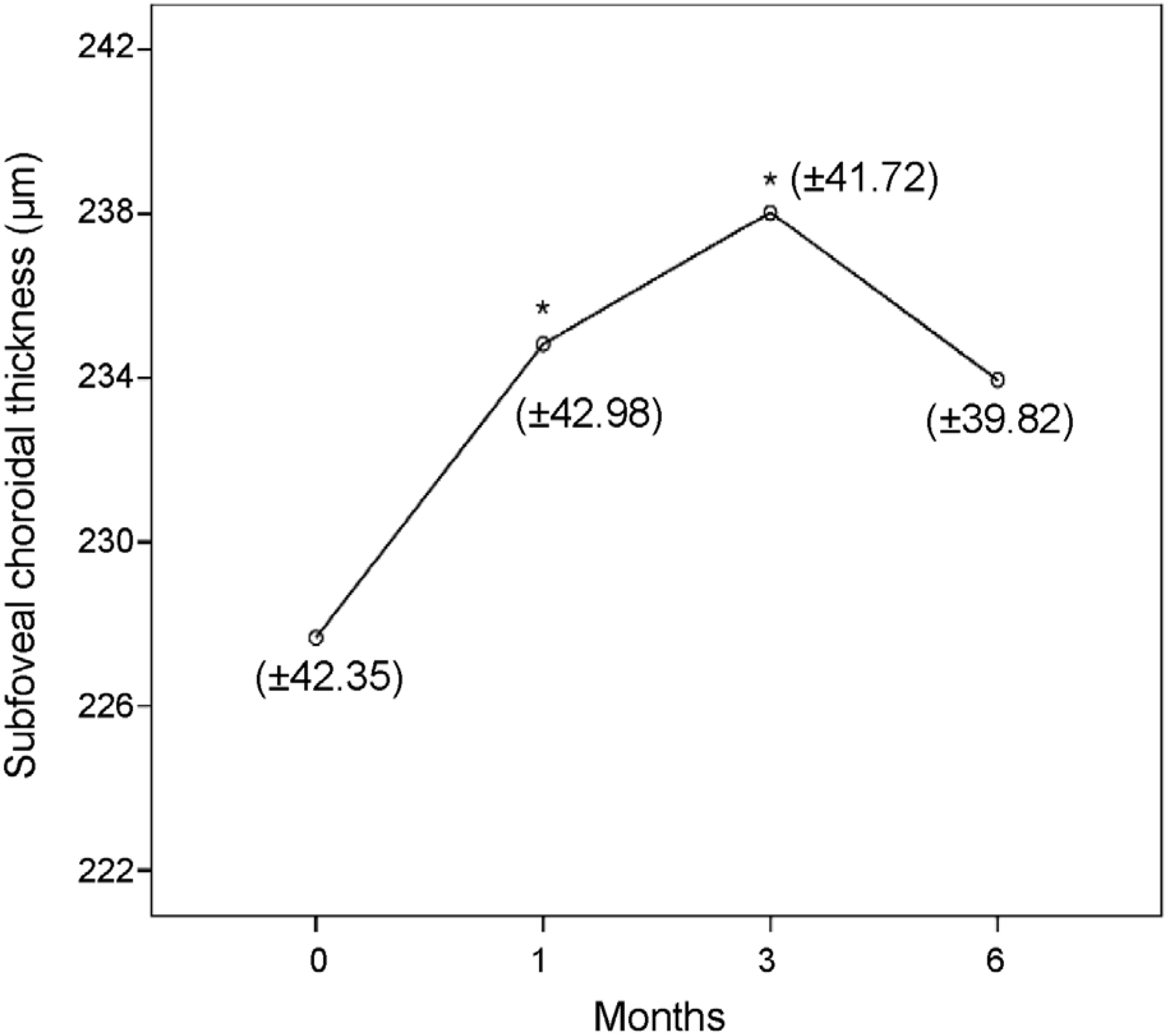 | Figure 1.Changes in subfoveal choroidal thickness after cataract surgery. The asterisks (*) indicate significant changes compared with preoperative values (paired t-test). |
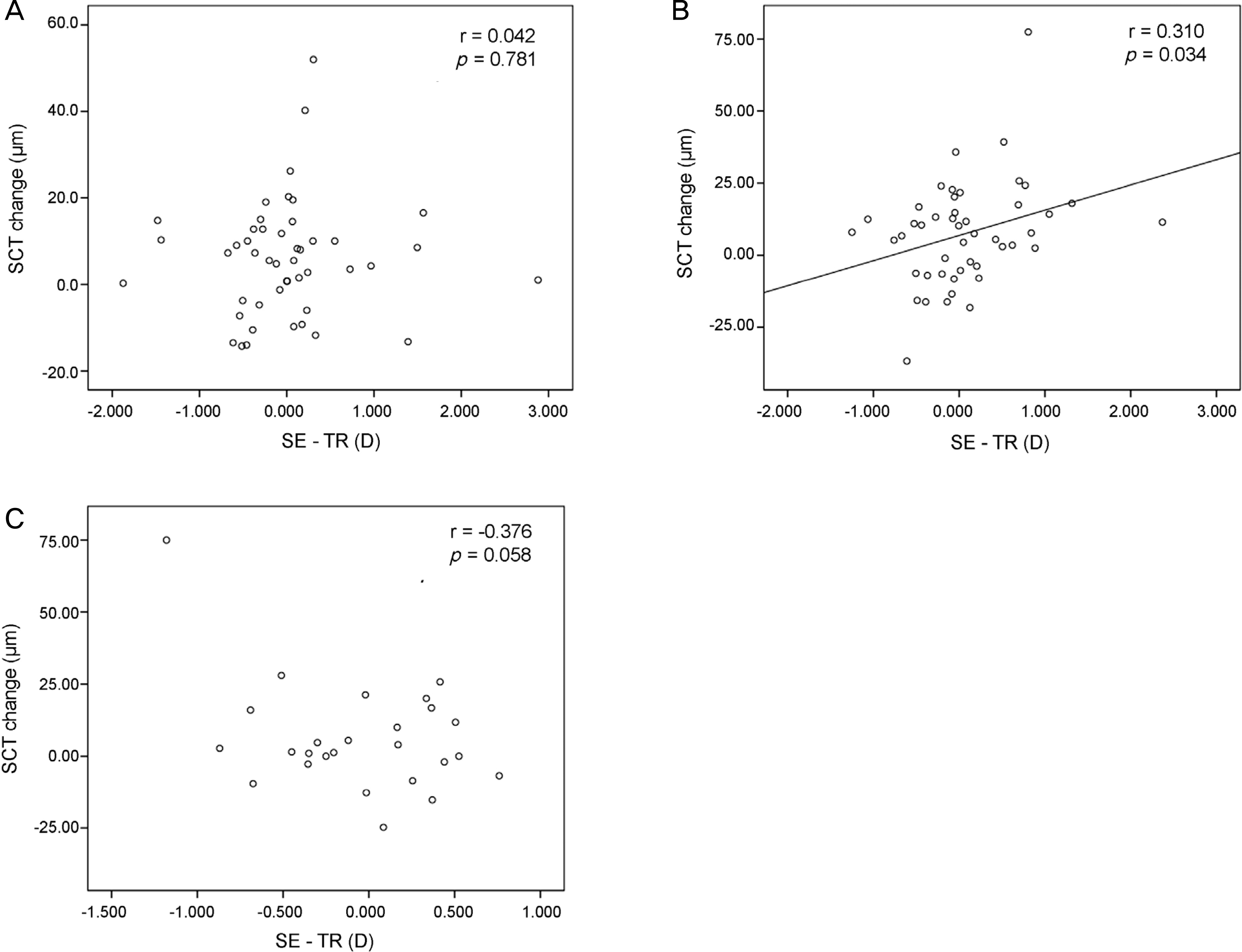 | Figure 2.Scatterplots showing the correlation between difference in the postoperative spherical equivalent (SE) and target refraction (TR) and the change in subfoveal choroidal thickness (SCT). (A) At 1 month after surgery. (B) At 3 months after surgery. (C) At 6 months after surgery. |
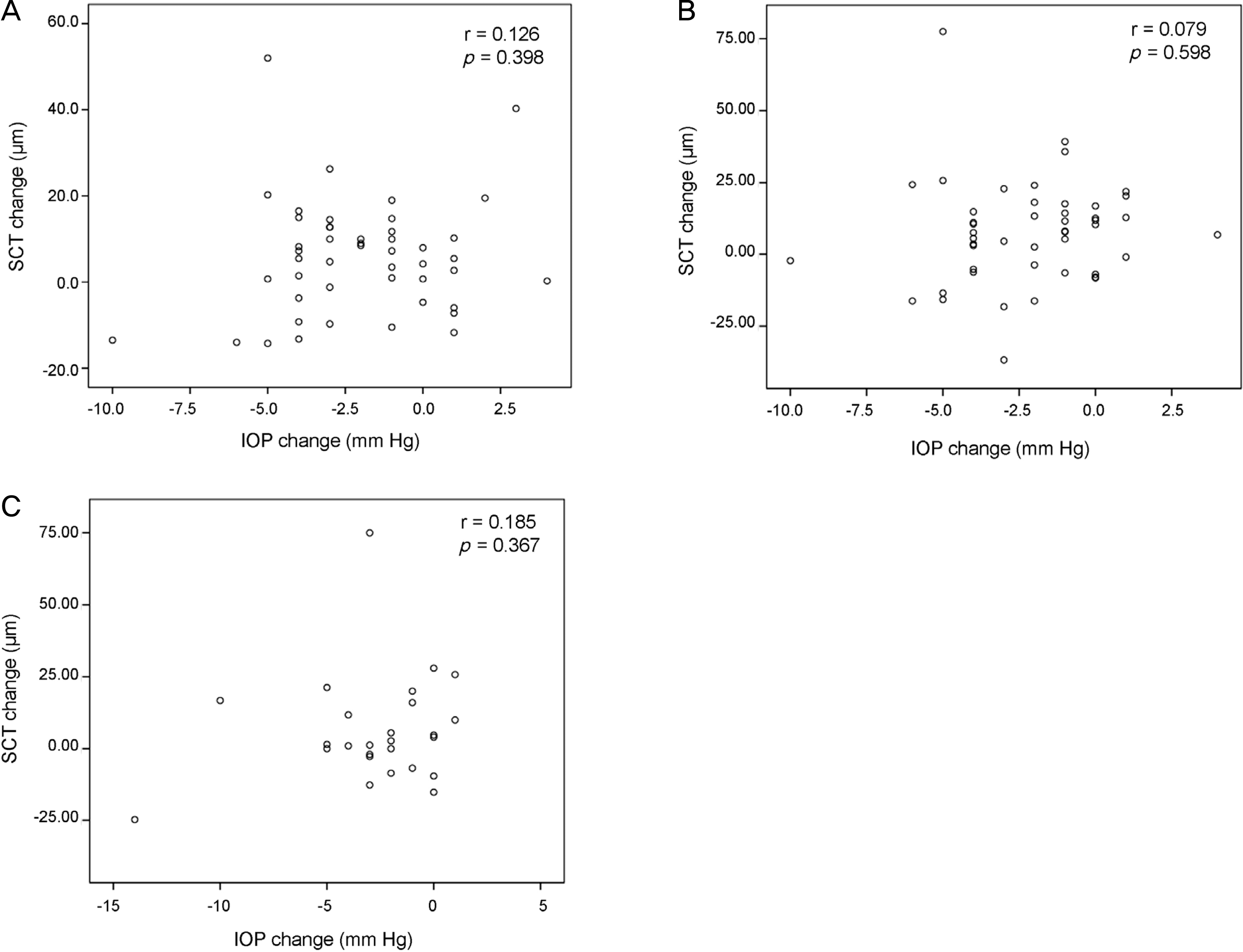 | Figure 3.Scatterplots showing the correlation between post-operative changes in intraocular pressure (IOP) and subfoveal choroidal thickness (SCT). (A) At 1 month after surgery. (B) At 3 months after surgery. (C) At 6 months after surgery. |
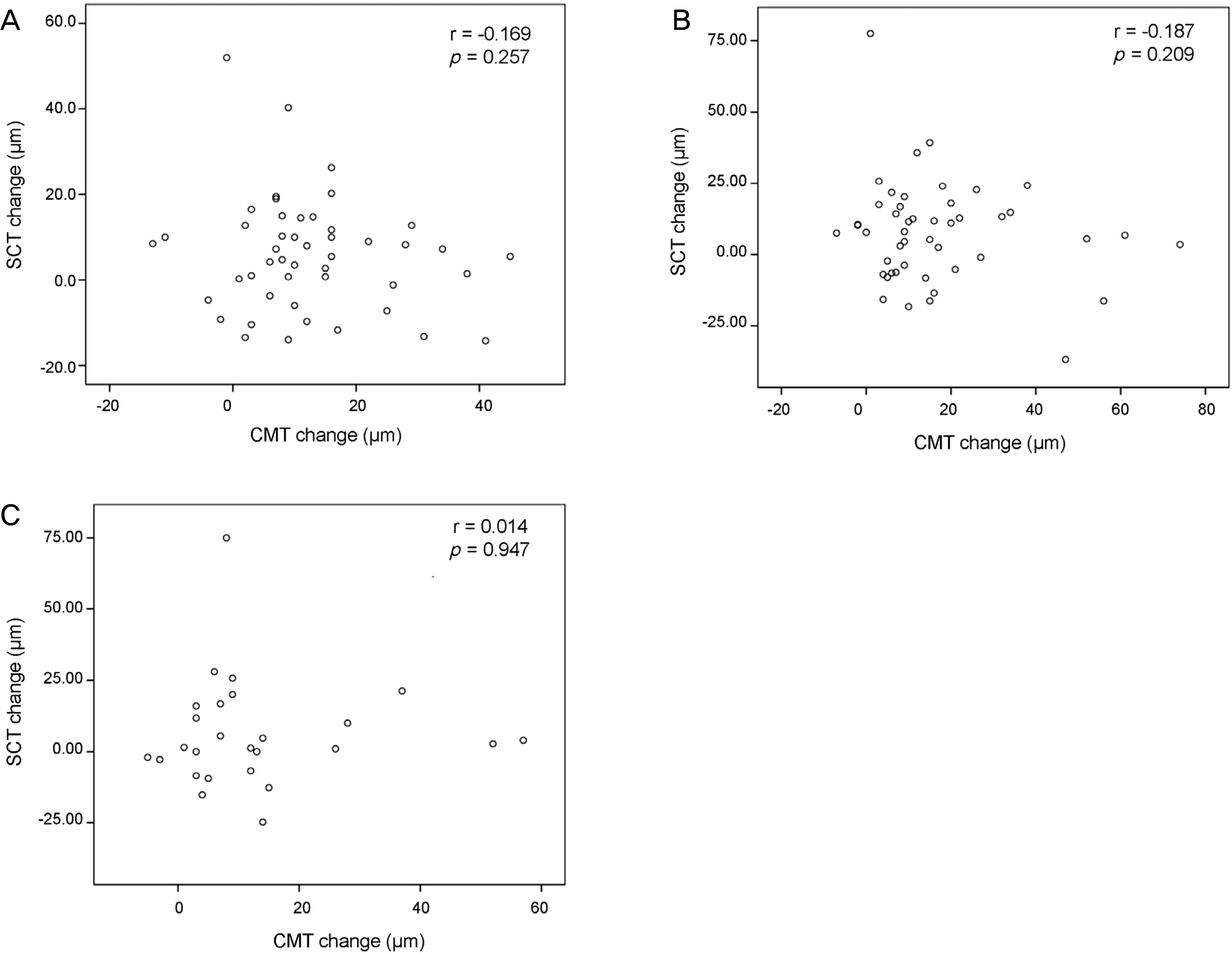 | Figure 4.Scatterplots showing the correlation between post-operative changes in central macular thickness (CMT) and subfoveal choroidal thickness (SCT). (A) At 1 month after surgery. (B) At 3 months after surgery. (C) At 6 months after surgery. |
Table 1.
Preoperative and postoperative measurements
| Parameter | Preoperative (n = 47) | 1 month (n = 47) | p-value* | 3 months (n = 47) | p-value* | 6 months (n = 26) | p-value* |
|---|---|---|---|---|---|---|---|
| SCT (μ m) | 228.7 ± 43.0 | 234.5 ± 41.7 | 0.004 | 236.2 ± 42.4 | 0.006 | 233.9 ± 39.8 | 0.104 |
| CMT (μ m) | 241.1 ± 21.1 | 253.7 ± 23.0 | <0.001 | 257.9 ± 24.9 | <0.001 | 249.9 ± 21.2 | <0.001 |
| IOP (mm Hg) | 12.8 ± 2.7 | 10.8 ± 2.3 | <0.001 | 10.6 ± 2.2 | <0.001 | 10.1 ± 2.6 | <0.001 |
 XML Download