

 PDF
PDF ePub
ePub Citation
 Print
Print


Abstract
Purpose
To evaluate the clinical results after phacoemulsification in mature and immature cataracts.
Methods
Mature cataract was defined as a classification of C5 by Lens Opacities Classification System III compared with other types of cataracts as controls. The present study included 37 (37 eyes) patients diagnosed with mature cataracts that received phacoemulsification and were followed up for at least 1 year. Thirty-seven (37 eyes) patients with other types of cataracts were selected randomly as controls. Intraoperative factors and rate of complications during and after surgery were evaluated. Best corrected visual acuity (BCVA), corneal endothelial cell density and central macular thickness (CMT) were measured during the 1 year of follow-up and compared with the controls.
Results
Twenty-seven eyes (mature cataracts) and 36 eyes (controls) received a complete continuous curvilinear capsulorhexis (CCC). The success rate of complete CCC was significantly high in the control eyes (p = 0.025). However, in mature cataract patients, 3 cases of posterior capsule rupture occurred among the 6 cases of radial tear of the anterior capsule, resulting in implantation of the lens in the sulcus. Posterior capsular ruptures were observed in 4 patients with mature cataracts and in 1 control with no statistically significant difference in the occurrence rate. Total phacoemulsification time and effective phacoemulsification time were significantly longer in the mature cataract patients (p = 0.038 and p = 0.041, respectively). BCVA, the amount of corneal endothelial cell density reduction and CMT at postoperative 1 year was not different between the two groups.
Go to : 
References
1. Shyn KH, Hong NS, Ahn SK. The prevalence and morphological characteristics of senile cataract in the local areas of Korea. J Korean Ophthalmol Soc. 1992; 33:1154–61.
2. Brusini P. Capsulorhexis in mature cataracts: why not? Doc Ophthalmol. 1992; 81:281–4.

3. Gimbel HV, Willerscheidt AB. What to do with limited view: the intumescent cataract. J Cataract Refract Surg. 1993; 19:657–61.
4. Mansour AM. Anterior capsulorhexis in hypermature cataracts. J Cataract Refract Surg. 1993; 19:116–7.
5. Vajpayee RB, Angra SK, Honavar SG, et al. Capsulotomy for abdominal in hypermature cataracts. J Cataract Refract Surg. 1995; 21:612–5.
6. Vasavada A, Singh R, Desai J. Phacoemulsification of white abdominal cataracts. J Cataract Refract Surg. 1998; 24:270–7.
7. Singh R, Vasavada AR, Janaswamy G. Phacoemulsification of bru-nescent and black cataracts. J Cataract Refract Surg. 2001; 27:1762–9.
8. Pande MV, Spalton DJ, Kerr-Muir MG, Marshall J. Postoperative inflammatory response to phacoemulsification and extracapsular cataract surgery: aqueous flare and cells. J Cataract Refract Surg. 1996; 22(Suppl 1):770–4.
9. Minassian DC, Rosen P, Dart JK, et al. Extracapsular cataract extraction compared with small incision surgery by abdominal: a randomised trial. Br J Ophthalmol. 2001; 85:822–9.
10. Melles GR, de Waard PW, Pameyer JH, Houdijn Beekhuis W. Trypan blue capsule staining to visualize the capsulorhexis in abdominal surgery. J Cataract Refract Surg. 1999; 25:7–9.
11. Chakrabarti A, Singh S. Phacoemulsification in eyes with white cataract. J Cataract Refract Surg. 2000; 26:1041–7.
12. Gimbel HV. Two-stage capsulorhexis for endocapsular phacoemulsification. J Cataract Refract Surg. 1990; 16:246–9.
13. Vasavada A, Singh R. Surgical techniques for difficult cataracts. Curr Opin Ophthalmol. 1999; 10:46–52.
14. Kim KB, Jeun EJ, Kim JC. Clinical results of the eyes with abdominal capsule rupture during cataract operation. J Korean Ophthalmol Soc. 1998; 39:2647–52.
15. Koo BS, Park YJ. Incidence of posterior capsular rupture in the learning curve of phacoemulsification. J Korean Ophthalmol Soc. 1994; 35:196–201.
16. Park YI, Oh TH, Choi KY. Clinical results of phacoemulsification performed by ophthalmic resident. J Korean Ophthalmol Soc. 1995; 36:1687–93.
17. Wright PL, Wilkinson CP, Balyeat HD, et al. Angiographic cystoid macular edema after posterior chamber lens implantation. Arch Ophthalmol. 1988; 106:740–4.
18. Henderson BA, Kim JY, Ament CS, et al. Clinical pseudophakic cystoid macular edema. Risk factors for development and duration after treatment. J Cataract Refract Surg. 2007; 33:1550–8.
19. Ursell PG, Spalton DJ, Whitcup SM, Nussenblatt RB. Cystoid macular edema after phacoemulsification: relationship to blood-aqueous barrier damage and visual acuity. J Cataract Refract Surg. 1999; 25:1492–7.
20. Flach AJ. The incidence, pathogenesis and treatment of cystoid macular edema following cataract surgery. Trans Am Ophthalmol Soc. 1998; 96:557–634.
21. Jacobson DR, Dellaporta A. Natural history of cystoid macular edema after cataract extraction. Am J Ophthalmol. 1974; 77:445–7.
22. Jampol LM, Sanders DR, Kraff MC. Prophylaxis and therapy of aphakic cystoid macular edema. Surv Ophthalmol. 1984; 28(Suppl):535–9.
Go to :
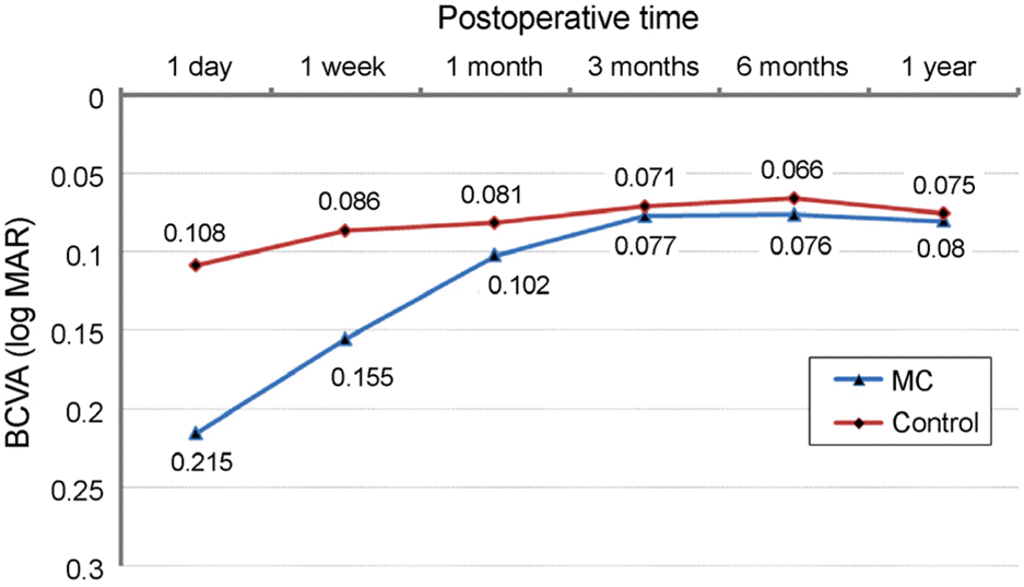 | Figure 1.Postoperative changes in best corrected visual acuity (BCVA) (log MAR). Although the difference in BCVA between mature cataract and control patients was significant on postoperative day 1 and week 1 (p = 0.031 and p = 0.04, respectively), the difference was not significant at the 1-year fol-low-up (p > 0.05). MC = mature cataract. |
Table 1.
Baseline demographic features of enrolled patients
| Mature cataract | Control | p-value | |
|---|---|---|---|
| Number (eyes) | 37 | 37 | |
| Age (year ± SD) | 74.8 ± 9.63 | 70.1 ± 11.76 | 0.79* |
| Gender (male/female) | 16/21 | 20/17 | |
| Preop BCVA (log MAR) | 0.910 ± 0.132 | 0.733 ± 0.085 | 0.31* |
| DM (%) | 35.1 | 40.5 | 0.82† |
| Dysthyroidism (%) | 2.78 | 0 | – |
Table 2.
Comparison of intraoperative and postoperative complications between the mature cataract and control patients
| Mature cataract (%) | Control (%) | p-value* | |
|---|---|---|---|
| Incomplete CCC | 10 (27.0) | 1 (2.7) | 0.025 |
| Posterior capsule rupture | 4 (10.8) | 1 (2.7) | 0.68 |
| Decentration of IOL optic | 4 (10.8) | 1 (2.7) | 0.68 |
Table 3.
Comparison of mean surgical parameters between the mature cataract and control patients
| Mature cataract | Control | p-value* | |
|---|---|---|---|
| Mean phaco time (sec) | 19.35 ± 9.26 | 13.53 ± 10.17 | 0.21 |
| Total phaco (%) | 7.53 ± 3.21 | 4.94 ± 3.08 | 0.038 |
| EPT (sec) | 4.19 ± 3.17 | 1.19 ± 1.16 | 0.041 |
| Total BSS used (mL) | 59.0 ± 10.02 | 51.8 ± 6.1 | 0.098 |
| Time of surgery (min) | 25.44 ± 8.99 | 20.66 ± 6.37 | 0.39 |
Table 4.
Postoperative changes in BCVA (log MAR), reduction of corneal endothelium and central foveal thickness
| Mature cataract | Control | p-value* | |
|---|---|---|---|
| Postop. BCVA | |||
| 1 day | 0.215 ± 0.061 | 0.108 ± 0.034 | 0.031 |
| 1 week | 0.155 ± 0.021 | 0.086 ± 0.029 | 0.04 |
| 1 month | 0.102 ± 0.019 | 0.081 ± 0.011 | 0.37 |
| 3 months | 0.077 ± 0.027 | 0.071 ± 0.013 | 0.56 |
| 6 months | 0.076 ± 0.018 | 0.066 ± 0.008 | 0.85 |
| 1 year | 0.080 ± 0.010 | 0.075 ± 0.012 | 0.76 |
| Postop. reduction of corneal endothelium (cells/mm2) | |||
| 1 month | 188 ± 89 | 150 ± 88 | 0.33 |
| 1 year | 190 ± 62 | 158 ± 42 | 0.35 |
| Central foveal thickness (μm) | |||
| Postop. 1 year | 201 ± 56 | 187 ± 71 | 0.63 |
 XML Download