Abstract
Purpose
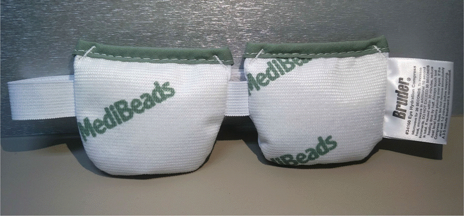
Warm compression using a commercial heat pad was used to evaluate the effects of temperature on the eyelids and tear film lipid layer thickness.
Methods
Targeting 13 patients (26 eyes) with non-specific eye disease such as dry eye syndrome or Meibomian gland dysfunction, we measured the average thickness of the tear film lipid layer in both eyes with the LipiView 2® System (Tearscience®, Morrisville, NY, USA). We performed warm compression on the right eye only in order to evaluate the effectiveness of massage and measured the temperature of the right eye lid immediately, 3 minutes, and 5 minutes after compression in order to compare with the untreated left eye. After warm compression for 5 minutes, we measured tear film lipid layer thickness of both eyes and analyzed the effectiveness of warm compression.
Results
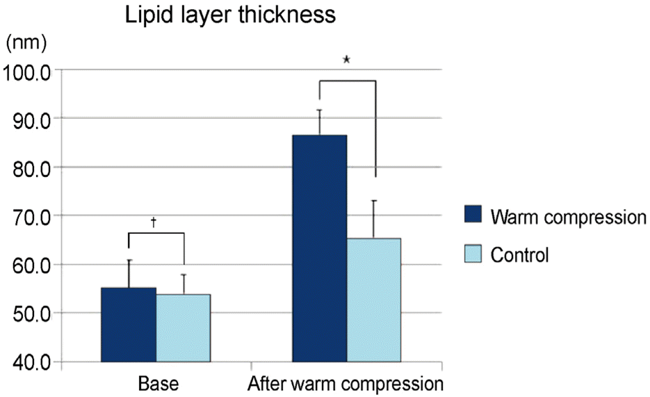
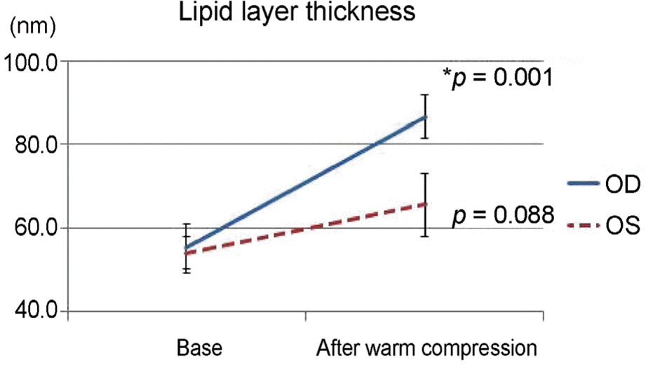
The average tear film lipid layer thickness was 55.1 ± 21.0 nm in the right eyes and 53.9 ± 13.9 nm in the left eyes (p = 0.474). Before performing the warm compression, the temperature of the right eye lid was 53.9 ± 13.9 nm, and that of the left was 35.9 ± 0.2 ° C. The eye lid temperature of the right eye immediately, 3 minutes, and 5 minutes after warm compression was 40.3 ± 1.3 ° C, 40.3 ± 1.3 ° C, and 40.3 ± 1.9 ° C, respectively, and these temperatures were relatively constant during the massage. Tear film lipid layer thickness after warm compression in the right eye was 83.5 ± 18.8 nm, which was increased compared to the original temperature (p = 0.001) and showed significant difference compared with the 65.5 ± 27.1 nm in the left eye (p = 0.005).
Go to : 
References
3. Mishima S, Maurice DM. The oily layer of the tear film and evaporation from the corneal surface. Exp Eye Res. 1961; 1:39–45.

4. Nelson JD, Shimazaki J, Benitez-del-Castillo JM, et al. The international workshop on meibomian gland dysfunction: report of the definition and classification subcommittee. Invest Ophthalmol Vis Sci. 2011; 52:1930–7.

5. Nichols KK, Foulks GN, Bron AJ, et al. The international abdominal on meibomian gland dysfunction: executive summary. Invest Ophthalmol Vis Sci. 2011; 52:1922–9.
6. Lemp MA. Report of the National Eye Institute/Industry workshop on clinical trials in dry eyes. CLAO J. 1995; 21:221–32.
7. Lee SH, Tseng SC. Rose bengal staining and cytologic abdominal associated with lipid tear deficiency. Am J Ophthalmol. 1997; 124:736–50.
8. Goto E, Monden Y, Takano Y, et al. Treatment of non-inflamed abdominal meibomian gland dysfunction by an infrared warm abdominal device. Br J Ophthalmol. 2002; 86:1403–7.
9. Ong BL, Larke JR. Meibomian gland dysfunction: some clinical, biochemical and physical observations. Ophthalmic Physiol Opt. 1990; 10:144–8.

10. Henriquez AS, Korb DR. Meibomian glands and contact lens wear. Br J Ophthalmol. 1981; 65:108–11.

11. Lee JK, Ha HS. Assessment of tear lipid layer after treatment by an infrared instrument. J Korean Ophthalmol Soc. 2004; 45:1659–64.
12. Petrofsky JS, Bains G, Raju C, et al. The effect of the moisture abdominal of a local heat source on the blood flow response of the skin. Arch Dermatol Res. 2009; 301:581–5.
Go to : 
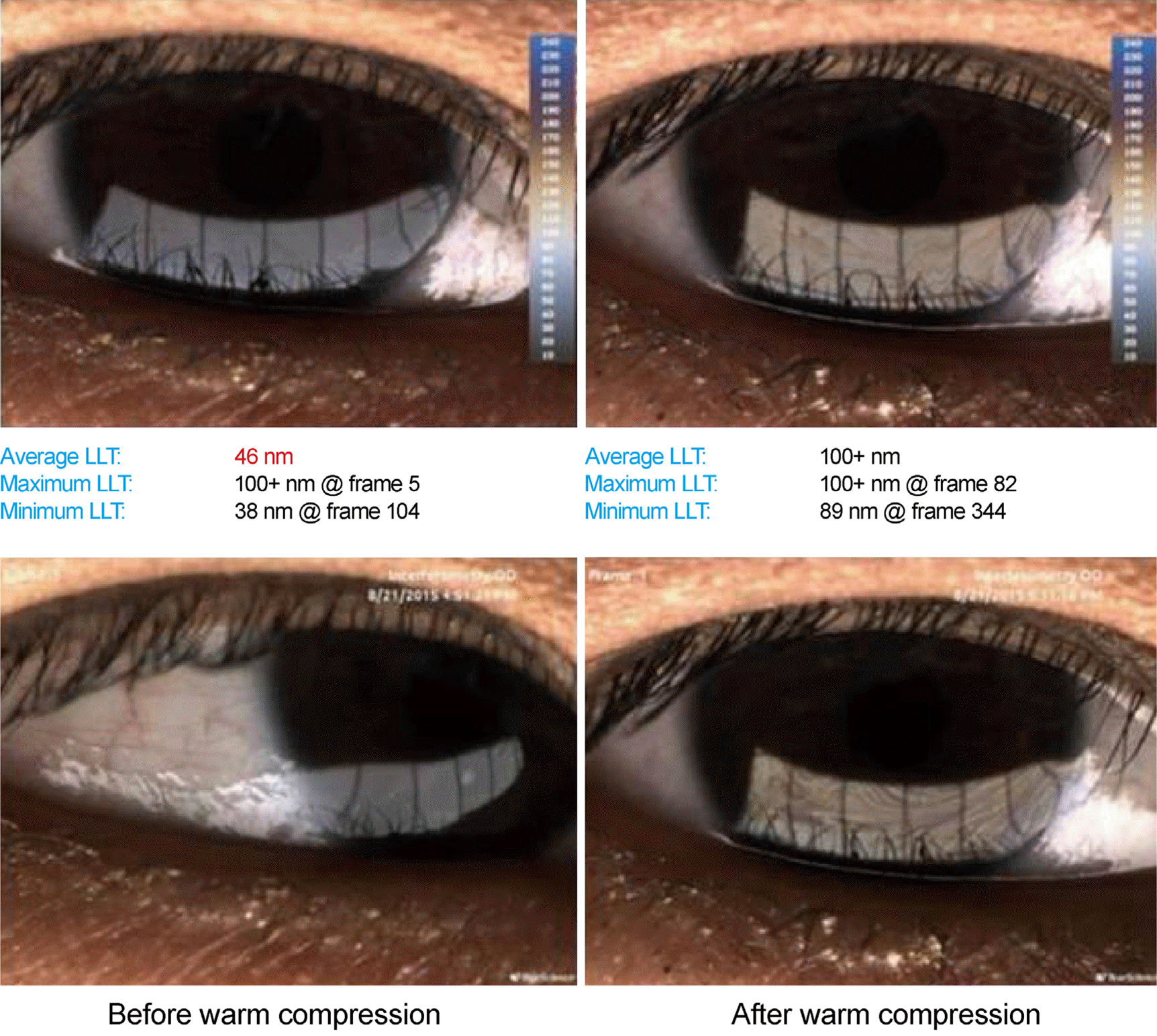
 | Figure 2.Example of average lipid layer thickness measured (LLT) by LipiView® System (Tearscience®, Morrisville, NY, USA). After warm compression, average LLT was increased 46 nm to 100 + nm. |
 | Figure 3.Comparison of average lipid layer thickness between warm compression eye and normal eye. p-value by Paired t-test. *p = 0.005; †p = 0.474. |
 | Figure 4.Comparison of average lipid layer thickness between before and after warm compression. OD = oculus dexter; OS = oculus sinister. * Paired t-test. |
Table 1.
Raw data of average lipid layer thickness of the normal group (Unit: nm)


 PDF
PDF ePub
ePub Citation
 Print
Print


 XML Download