

 PDF
PDF ePub
ePub Citation
 Print
Print


Abstract
Purpose
To report a case of an 82-year-old male with acute myeloid leukemia presenting with bilateral isolated conjunctival and eyelid masses.
Case summary
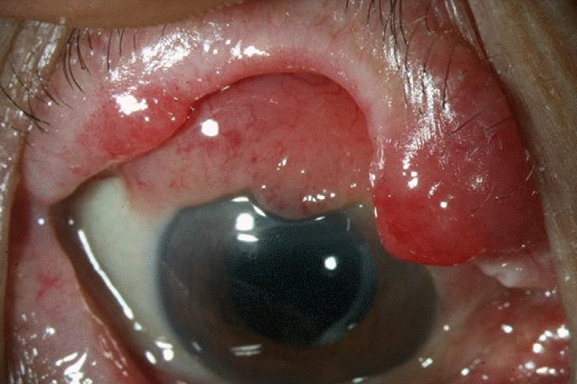
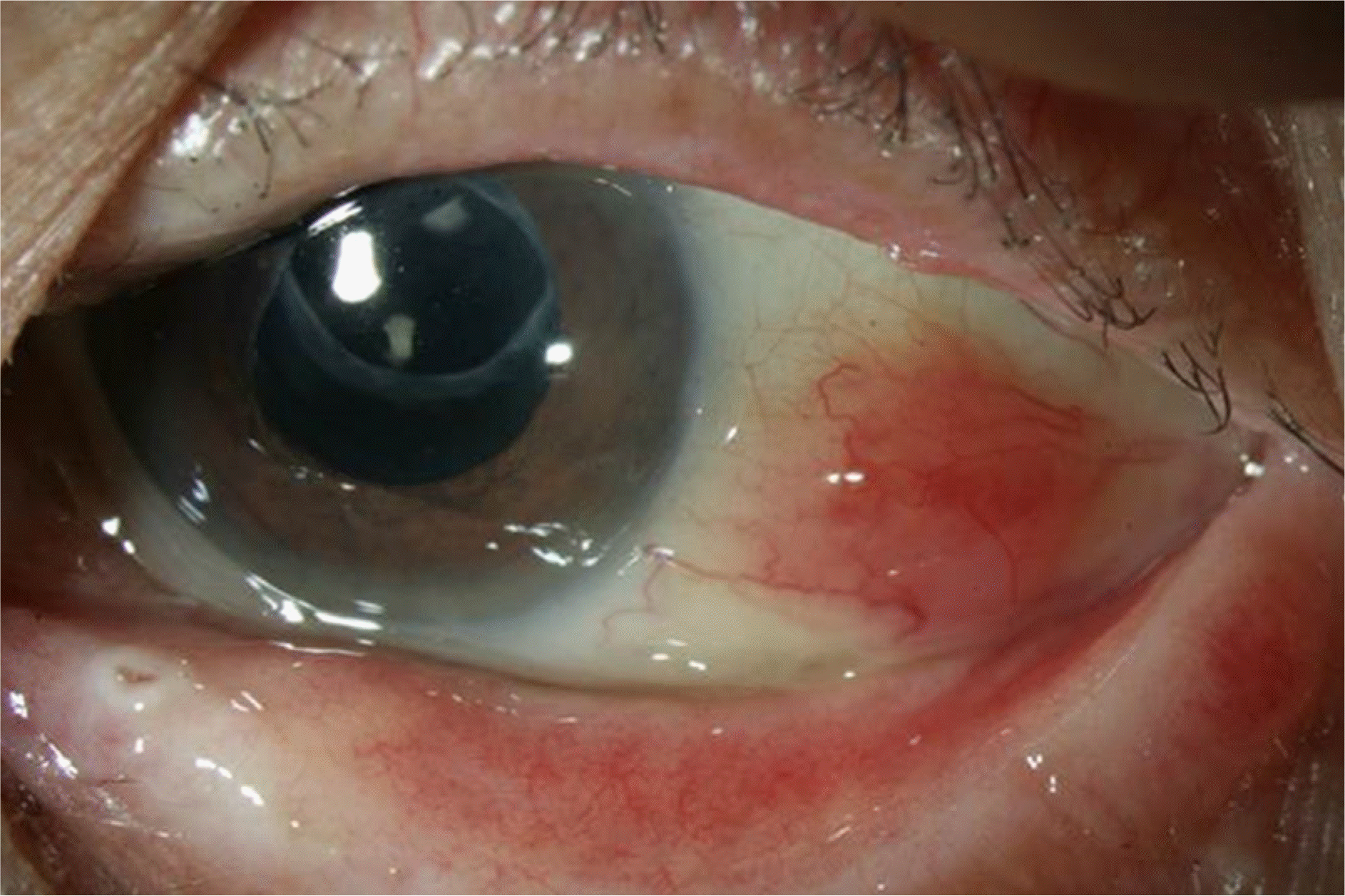
An 82-year-old male presented with a bilateral conjunctival mass and right eyelid mass occurring 10 days prior. He was diagnosed with prostate cancer 8 years ago and complete recovery was achieved using selective mass chemotherapy. He experienced a stroke 4 years ago and was treated using a carotid artery stent insertion and medication. In the initial laboratory test, hemoglobin was 13.7 g/dL and leukocyte count 5,530/mm3 (neutrophil 74.4%, lymphocyte 10%, monocyte 11.8%). Light reflex, movement of extraocular muscle and fundus examination were all normal. Biopsy was performed 1 week after the first visit. Seven days after biopsy, he complained of sudden dyspnea and febrile sense and was admitted to the intensive care unit via the emergency room (ER). The laboratory tests performed in the ER showed hemoglobin was 9.6 g/dL and leukocyte count was 78,020/mm3 (neutrophil 0%, lymphocyte 7%, monocyte 5%, promyelocyte 1%, metamyelocyte 4%, myelocyte 6%, blast 67%). The biopsy revealed diffuse proliferation of atypical plasmacytoid cells, consistent with leukemic infiltration. Under the diagnosis of acute myeloid leukemia, chemotherapy was administered. However, the patient died due to aggravated pneumonia.
References
1. Neiman RS. Barcos M. Berard C, et al. Granulocytic sarcoma: a clinicopathologic study of 61 biopsied cases. Cancer. 1981; 48:1426–37.

2. Sharma T. Grewal J. Gupta S. Murray PI. Ophthalmic manifestations of acute leukaemias: the ophthalmologist’s role. Eye (Lond). 2004; 18:663–72.
3. Charif Chefchaouni M. Belmekki M. Hajji Z, et al. Ophthalmic manifestations of acute leukemia. J Fr Ophtalmol. 2002; 25:62–6.
4. Brownstein S. Thelmo W. Olivier A. Granulocytic sarcoma of the orbit. Report of a case. Can J Ophthalmol. 1975; 10:174–83.
5. Méndez-Cepeda P. Millán-Rodríguez AC. Dios E, et al. Conjunctival myeloid sarcoma in acute myeloblastic leukemia-M1. Arch Soc Esp Oftalmol. 2012; 87:79–81.
6. Rosenberg C. Finger PT. Furlan L. Iacob CE. Bilateral epibulbar granulocytic sarcomas: a case of an 8-year-old girl with acute myeloid leukaemia. Graefes Arch Clin Exp Ophthalmol. 2007; 245:170–2.
7. Shields JA. Stopyra GA. Marr BP, et al. Bilateral orbital myeloid sarcoma as initial sign of acute myeloid leukemia: case report and review of the literature. Arch Ophthalmol. 2003; 121:138–42.
8. Kincaid MC. Green WR. Ocular and orbital involvement in leukemia. Surv Ophthalmol. 1983; 27:211–32.
9. Lee HS. Park JW. Yang SW. A case of granulocytic sarcoma involving the forniceal conjunctiva. J Korean Ophthalmol Soc. 2006; 47:986–90.

 XML Download