Abstract
Purpose
To report the clinical characteristics of retinoblastoma patients whose diagnosis was difficult due to atypical ocular manifestations.
Methods
Among retinoblastoma patients who were diagnosed and treated from January 1999 to December 2014 at Seoul National University Children’s Hospital, 6 patients whose diagnosis was difficult were retrospectively reviewed. Factors including age, sex, family history, initial findings, time to final diagnosis, histopathologic examination, additional treatment, and survival rate were evaluated.
Results
Among 6 patients, 5 were male, and the mean age at the initial visit was 32.9 ± 19.1 months. None of the patients had family history, and all presented with unilateral lesion at the initial visit. The initial diagnoses were Coats’ disease and uveitis in 2 patients, respectively, and persistent hyperplastic primary vitreous and traumatic hyphema in 1 patient, respectively. During an intensive short-term fdlow-up of 8.3 ± 5.3 weeks, 2 patients showed malignant cells after external subretinal fluid drainage procedure, and 4 patients demonstrated increasing ocular size or calcification in imaging. These patients received enucleation under suspicion of malignancy and were finally diagnosed with retinoblastoma after histopathologic examination. There were 2 patients with optic nerve involvement, and 3 patients underwent additional systemic chemotherapy. Five patients were followed-up for 7.6 ± 6.3 years after enucleation, and the mean age at final fdlow-up was 10.6 ± 7.4 years.
Conclusions
Retinoblastoma is one of the diseases in which early diagnosis and treatment are important. However, some cases are difficult to diagnose, even for experienced clinicians. If there are no typical manifestations such as mass or calcification and early findings show retinal detachment, glaucoma, pseudohypopyon, or hyphema, intensive short-term fdlow-up to exclude retinoblastoma is needed.
Go to : 
References
1. Ellsworth RM. The practical management of retinoblastoma. Trans Am Ophthalmol Soc. 1969; 67:462–534.
2. Seregard S. Lundell G. Svedberg H. Kivelä T. Incidence of retinoblastoma from 1958 to 1998 in Northern Europe: advantages of birth cohort analysis. Ophthalmology. 2004; 111:1228–32.
3. Broaddus E. Topham A. Singh AD. Incidence of retinoblastoma in the USA: 1975-2004. Br J Ophthalmol. 2009; 93:21–3.

5. Haider S. Qureshi W. Ali A. Leukocoria in children. J Pediatr Ophthalmol Strabismus. 2008; 45:179–80.

6. Stafford WR. Yanoff M. Parnell BL. Retinoblastomas initially misdiagnosed as primary ocular inflammations. Arch Ophthalmol. 1969; 82:771–3.

7. Shields JA. Shields CL. Differentiation of coats’ disease and retinoblastoma. J Pediatr Ophthalmol Strabismus. 2001; 38:262–6. quiz. 302–3.

8. Shields JA. Shields CL. Parsons HM. Differential diagnosis of retinoblastoma. Retina. 1991; 11:232–43.

9. Rodrigues KE. Latorre Mdo R. de Camargo B. Delayed diagnosisin retinoblastoma. J Pediatr (Rio J). 2004; 80:511–6.
10. Kim JH. Yu YS. Incidence (1991~1993) and survival rates (1991-2003) of retinoblastoma in Korea. J Korean Ophthalmol Soc. 2010; 51:542–51.

11. Patelli F. Zumbo G. Fasolino G, et al. Treatment and outcome of exudative retinal detachment in Coats disease: a case report. Semin Ophthalmol. 2004; 19:117–8.

14. Choi SY. Yu YS. Treatment and clinical results of Coats’ disease. J Korean Ophthalmol Soc. 1999; 40:2190–7.
15. Shields JA. Shields CL. Honavar SG, et al. Classification and management of Coats disease: the 2000 Proctor Lecture. Am J Ophthalmol. 2001; 131:572–83.

16. Ma DJ. Choi J. Jang JW, et al. Bilateral Coats’ disease: a case report. J Korean Ophthalmol Soc. 2011; 52:112–6.

17. Miller DM. Benz MS. Murray TG. Dubovy SR. Intraretinal calcification and osseous metaplasia in coats disease. Arch Ophthalmol. 2004; 122:1710–2.

18. Lam HD. Samuel MA. Rao NA. Murphree AL. Retinoblastoma presenting as Coats’ disease. Eye (Lond). 2008; 22:1196–7.

19. Shields CL. Uysal Y. Benevides R, et al. Retinoblastoma in an eye with features of Coats’ disease. J Pediatr Ophthalmol Strabismus. 2006; 43:313–5.

20. Jack RL. Regression of the hyaloid vascular system. An ultrastructural analysis. Am J Ophthalmol. 1972; 74:261–72.
21. Kyung HS. Yu YS. Clinical findings and prognosis of persistent hyperplastic primary vitreous. J Korean Ophthalmol Soc. 2004; 45:1528–34.
22. Català-Mora J. Parareda-Salles A. Vicuña-Muñoz CG, et al. Uveitis masquerade syndrome as a presenting form of diffuse retinoblastoma. Arch Soc Esp Oftalmol. 2009; 84:477–80.
Go to : 
 | Figure 1.Imaging of patient 2. (A) Initial fundus photograph shows serous bullous kissing type retinal detachment with white opaque particles in the left eye. (B) Initial B-scan of the left eye shows increased echogenicity in the vitreous cavity, without calcification. (C) Initial computed tomography shows mild increased density in the left eye without evidence of a mass or calcification. (D) Two months after the initial visit, heterogeneous signal intensity in the left eye was noted on magnetic resonance imaging. |
 | Figure 2.Imaging of patient 3. (A) Initial B-scan shows no definite mass-like lesion or calcification in the left eye. (B) Anterior segment photograph 2 months after Ahmed valve implantation shows an edematous cornea, shallow anterior chamber, and ciliary injection in the left eye. |
 | Figure 3.Imaging of patient 5. (A) Initial anterior segment photograph of the right eye shows hypopyon and whitish spots on the surface of the iris. (B-D) There is no evidence of mass-like lesion or calcification on B-scan, computed tomography, or magnetic resonance imaging of the right eye at the initial visit. |
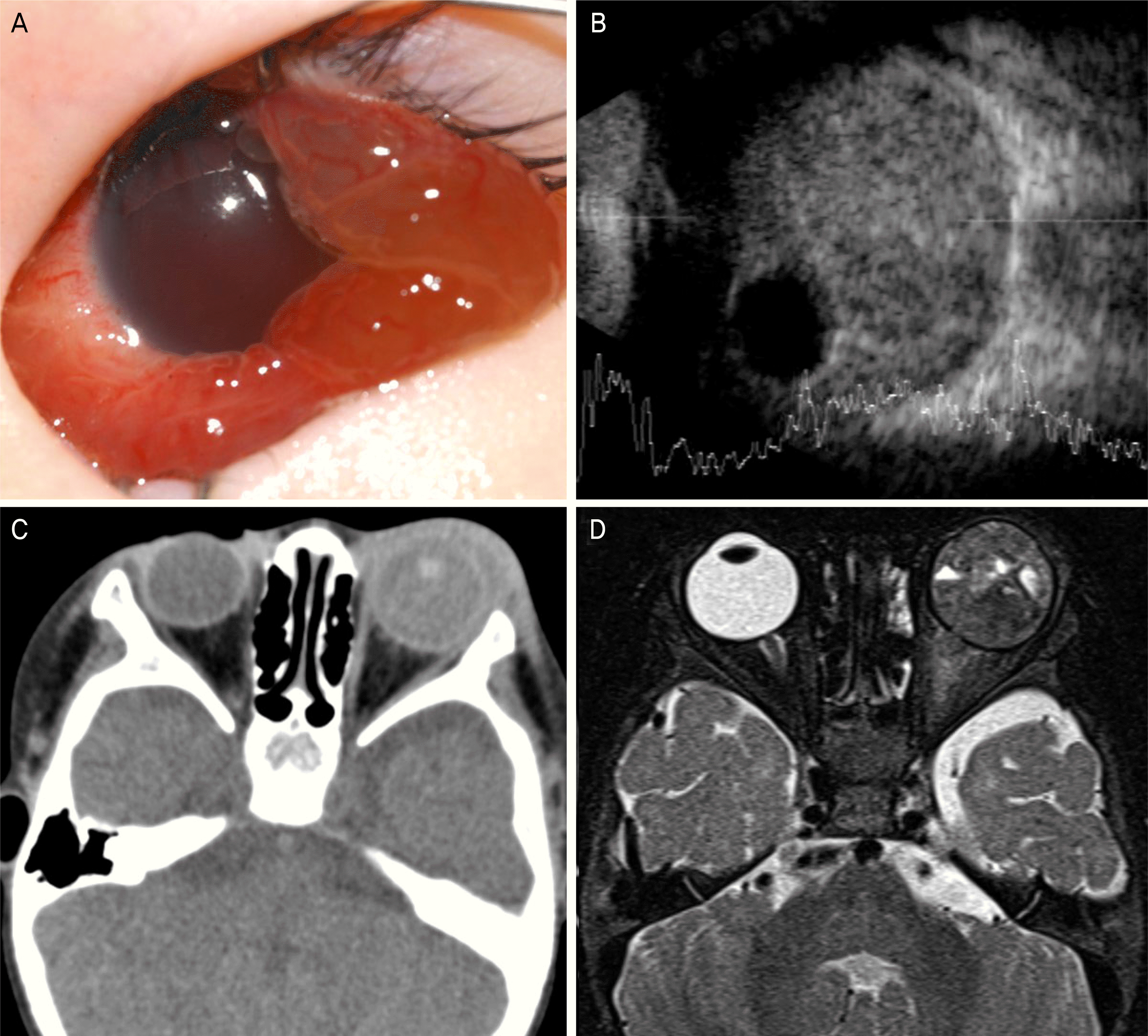
 | Figure 4.Imaging of patient 6. (A) Anterior segment photograph of the left eye at the initial visit shows diffuse chemosis and hyphema. (B, C) Initial B-scan of the left eye and computed tomography show increased echogenicity and a dislocated lens in the vitreous cavity without evidence of mass-like lesion or calcification. (D) After 2.5 months from the initial visit, heterogeneous signal intensity in the left eye was suspicious for retinoblastoma on magnetic resonance imaging. |
Table 1.
Clinical characteristics of patients with retinoblastoma at initial diagnosis
R = right; L = left; CT = computed tomography; M = male; F = female; RD = retinal detachment; SRFD = subretinal fluid drainage; NVI = neovascularizqtion of the iris; VH = vitreous hemorrhage; PHPV = persistent hyperplastic primary vitreous; VT = vitrectomy; AGV = ahmed glaucoma valve; TA = triamcinolone acetonide; AC = anterior chamber.
Table 2.
Clinical characteristics of patients with retinoblastoma at the final diagnosis and during the follow-up period


 PDF
PDF ePub
ePub Citation
 Print
Print


 XML Download