Abstract
Purpose
Isolated inferior oblique weakening procedure is an effective treatment for patients with superior oblique muscle palsy who had up to 15 prism diopters (PD) of vertical deviation in the primary position, but 2-muscle surgery is needed for patients with larger deviations. Herein, we report the surgical results of simultaneous 2-extraocular muscle surgery for large primary position hypertropia 16 PD or more caused by superior oblique palsy.
Methods
This study was a retrospective review of the records of patients who presented with central gaze hypertropia 16 PD or more and underwent simultaneous 2-extraocular muscle surgery between January 2003 and June 2014 in Severance Hospital. The patients were divided into 3 groups: 43 patients who underwent inferior oblique (IO) myectomy and contralateral inferior rectus (IR) recession (Group 1), 10 patients who underwent IO myectomy and superior rectus (SR) recession (Group 2), and 8 patients who underwent SR recession and contralateral IR recession (Group 3). Criteria for success included correction of head posture and a primary position alignment within 5 PD of vertical deviation.
Results
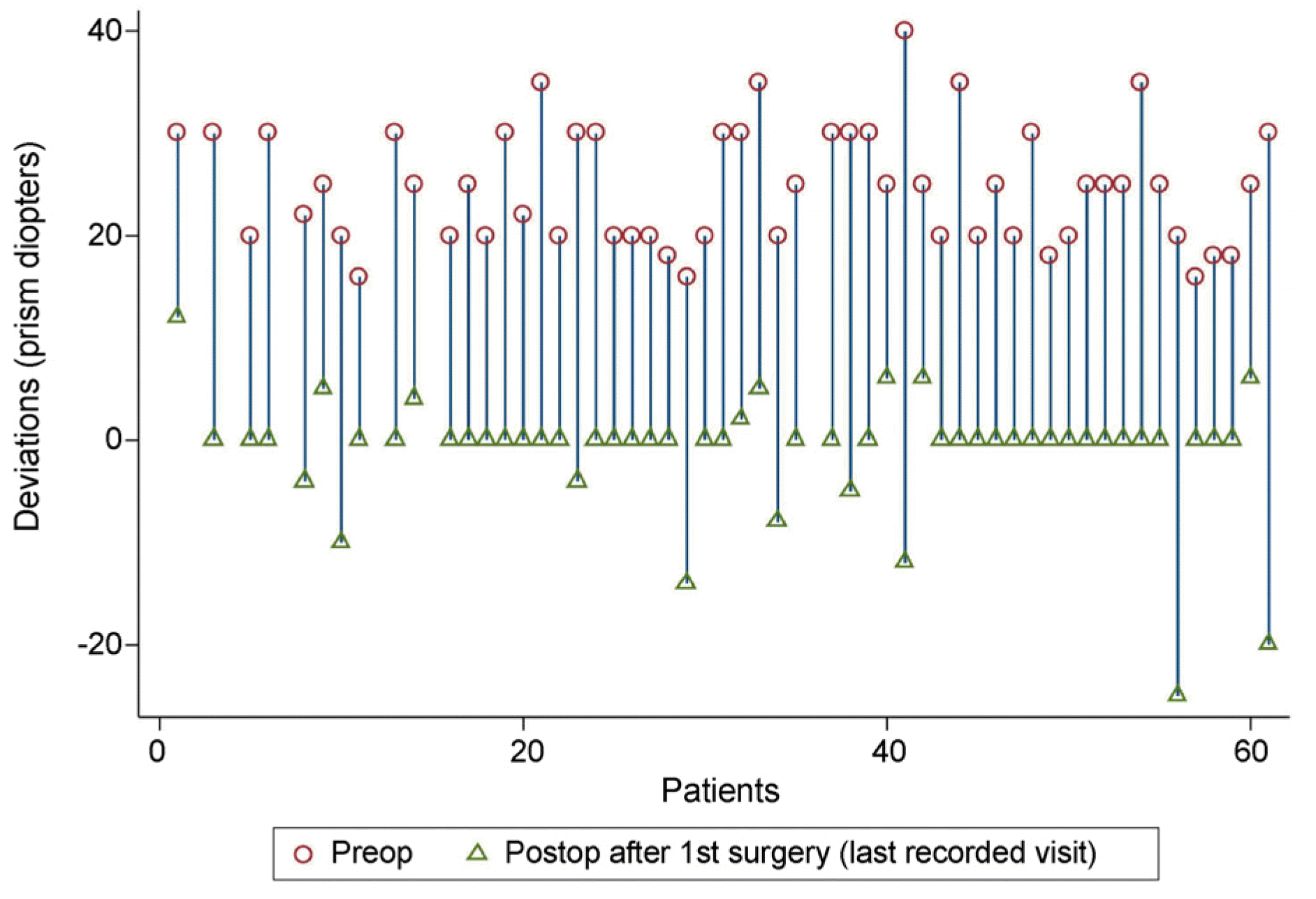
Mean preoperative alignment at primary gaze was 25.5 ± 7.1 PD (range, 16-60 PD) compared to the postoperative value of -1.3 ± 6.8 PD (range, -20~25 PD) (p < 0.001). Surgery was successful in 49 (80%) patients. Nine (15%) patients were overcorrected and the other 3 (5%) patients were undercorrected. Success rate was the highest in subjects who underwent IO myectomy and contralateral IR recession. Among the 24 patients who did not receive combined horizontal muscle surgery, horizontal deviations decreased from 10.4 ± 2.7 PD to 1.5 ± 5.5 PD (p < 0.001)
Conclusions
Two-muscle surgery can be effective in patients with large hypertropia 16 PD or more. Additionally, horizontal deviations are more likely to be resolved with vertical muscle surgery alone. However, IO myectomy combined with ipsilateral SR recession can cause overcorrection postoperatively, so surgical dose should be reduced when performing weakening procedure of two elevators in one eye.
References
1. Tamhankar MA. Kim JH. Ying GS. Volpe NJ. Adult hypertropia: a guide to diagnostic evaluation based on review of300 patients. Eye (Lond). 2011; 25:91–6.
2. Tollefson MM. Mohney BG. Diehl NN. Burke JP. Incidence and types of childhood hypertropia: a population-based study. Ophthalmology. 2006; 113:1142–5.
3. Saunders RA. Treatment of superior oblique palsy with superior oblique tendon tuck and inferior oblique muscle myectomy. Ophthalmology. 1986; 93:1023–7.

4. Morad Y. Weinstock VM. Kraft SP. Outcome of inferior oblique recession with or without vertical rectus recession for unilateral superior oblique paresis. Binocul Vis Strabismus Q. 2001; 16:23–8.
5. Madigan WP. Reynolds JD. Strominger M. Wagner RS. Management of congenital fourth cranial nerve palsy. J Pediatr Ophthalmol Strabismus. 2014; 51:70–2.

6. Shipman T. Burke J. Unilateral inferior oblique muscle myectomy and recession in the treatment of inferior oblique muscle overaction: a longitudinal study. Eye (Lond). 2003; 17:1013–8.

7. Nejad M. Thacker N. Velez FG, et al. Surgical results of patients with unilateral superior oblique palsy presenting with large hypertropias. J Pediatr Ophthalmol Strabismus. 2013; 50:44–52.

8. Knapp P. Moore S. Diagnosis and surgical options in superior oblique surgery. Int Ophthalmol Clin. 1976; 16:137–49.

9. von Noorden GK. Murray E. Wong SY. Superior oblique paralysis. A review of 270 cases. Arch Ophthalmol. 1986; 104:1771–6.
10. Hatz KB. Brodsky MC. Killer HE. When is isolated inferior oblique muscle surgery an appropriate treatment for superior oblique palsy? Eur J Ophthalmol. 2006; 16:10–6.

12. Ahn SJ. Choi J. Kim SJ. Yu YS. Superior rectus muscle recession for residual head tilt after inferior oblique muscle weakening in superior oblique palsy. Korean J Ophthalmol. 2012; 26:285–9.

13. Wright KW. Late overcorrection after inferior rectus recession. Ophthalmology. 1996; 103:1503–7.

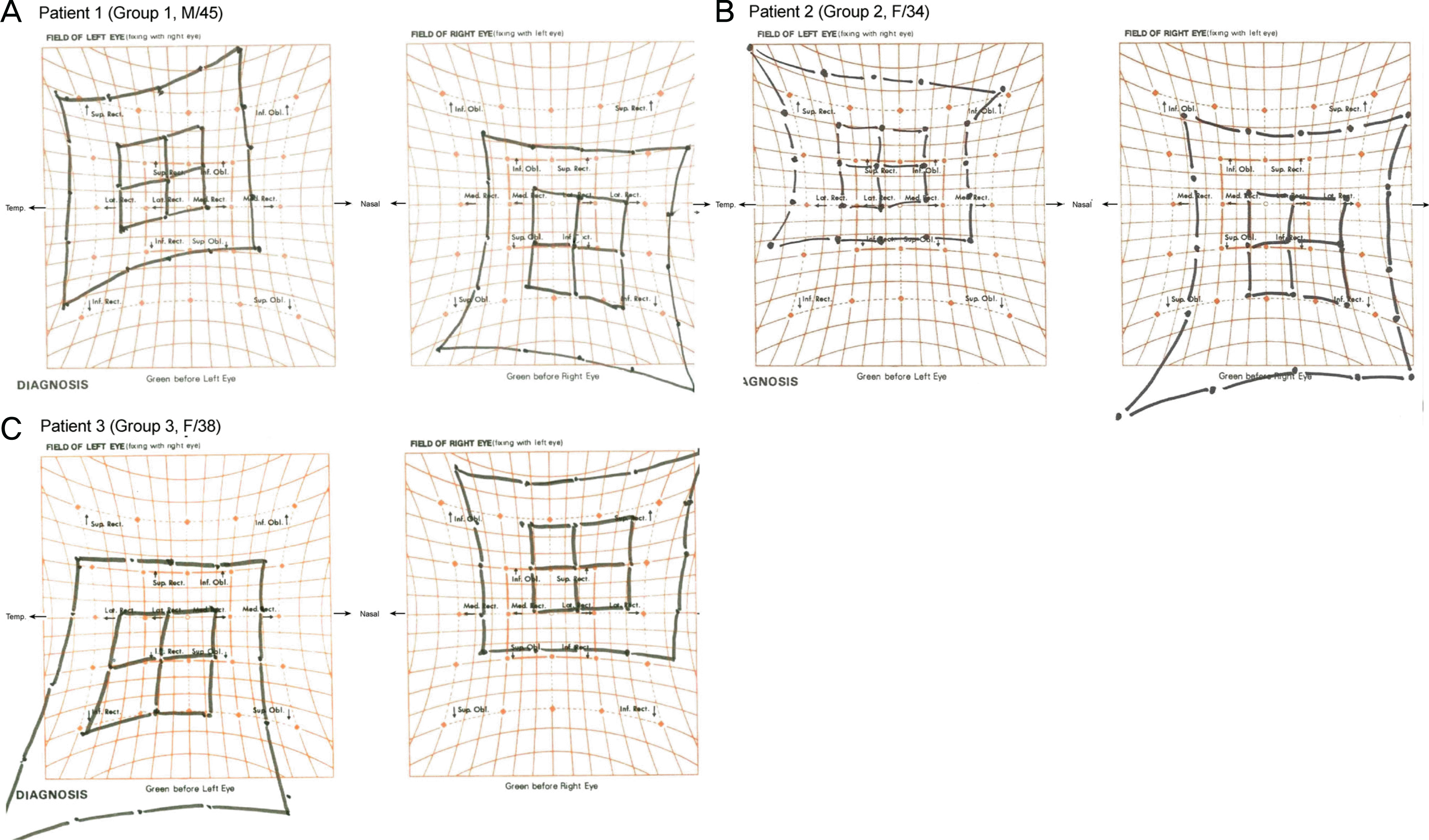
Figure 1.
Preoperative Hess screening tests of patients. (A) Group 1. A 45-year-old male with 18 PD exotropia and 30 PD left hypertropia at primary position. Duction and version testing revealed left inferior oblique overaction 3 + and left superior oblique underaction 2-. Left inferior oblique (LIO) myectomy and right inferior rectus (RIR) recession 5.0 mm was performed. (B) Group 2. A 34-year-old female with 12 PD exotropia and 30 PD left hypertropia at primary position with left inferior oblique overaction 3 + and right superior oblique overaction 4+. LIO myectomy and left superior rectus (LSR) recession 7.5 mm was performed in this patient. (C) Group 3. A 38-year-old female with 10 PD exotropia and 30 PD right hypertropia at primary position showed spread of comitance with no definite inferior oblique overaction. Right superior rectus (RSR) recession 10.0 mm and left inferior rectus (LIR) recession 5.5 mm was performed. PD = prism diopters; Sup = superior; Rect = rectus; Inf = inferior; Obl = oblique; Temp = temporal; Lat = lateral; Med = medial.

Figure 2.
The change of vertical deviations before and after simultaneous 2-muscle surgery in patients with large-angle (≥16 PD) superior oblique palsy. The graphs showed that overcorrection was observed in 9 patients (15%) after simultaneous 2-muscle surgery. Preop = preoperation; Postop = postoperation.

Table 1.
Patient characteristics
Table 2.
Clinical characteristics and surgical outcomes according to the first procedure performed
| IO myectomy + contralateral IR recession (n = 43) | IO myectomy + SR recession (n = 10) | SR recession + contralateral IR recession (n = 8) | p-value | |
|---|---|---|---|---|
| Age at surgery (years) | 20.1 ± 16.9 (range, 2.1-78.8) | 17.5 ± 14.2 (range, 3.7-47.6) | 32.3 ± 20.5 (range, 8.5-72) | 0.093∗ |
| Hypertropia at primary (PD) | 26.3 ± 7.4 (range, 16.0-60.0) | 23.6 ± 7.0 (range, 16.0-40.0) | 23.6 ± 5.2 (range, 16.0-30.0) | 0.424† |
| Hypertropia at adduction (PD) | 29.1 ± 7.9 (range, 20.0-55.0) | 27.1 ± 8.5 (range, 16.0-40.0) | 23.5 ± 4.7 (range, 18.0-30.0) | 0.178† |
| Hypertropia at abduction (PD) | 16.6 ± 9.0 (range, 0.0-35.0) | 14.5 ± 10.1 (range, 0.0-30.0) | 18.6 ± 6.2 (range, 12.0-30.0) | 0.649† |
| Excyclotropia (°) | 4.7 ± 4.0 (range, 0.0-20.0) | 4.2 ± 4.9 (range, 0.0-10.0) | 4.4 ± 3.2 (range, 0.0-10.0) | 0.937† |
| Diplopia (n) | 6 (13.95%) | 1 (10%) | 2 (25%) | 0.725‡ |
| Adjustable suture (n) | 20 (46.5%) | 5 (50%) | 6 (75%) | 0.411‡ |
| Combined LR recession (n) | 8 (18.6%) | 1 (10.0%) | 3 | - |
| Successful outcome (n) | 37 (86.1%) | 6 (60%) | 6 (75%) | 0.131‡ |


 PDF
PDF ePub
ePub Citation
 Print
Print


 XML Download