Abstract
Purpose
To investigate the surgical outcome of Ahmed glaucoma valve implantation (AVI) combined with 23-gauge vitrectomy in eyes with medically uncontrolled neovascular glaucoma (NVG).
Methods
Thirty six eyes of 35 NVG patients who underwent AVI combined with 23-gauge vitrectomy and have been followed-up at least 6 months after surgery and were retrospectively reviewed. Surgical success was defined as the control of intraocular pressure (IOP) between 6 and 21 mm Hg, irrespective of the use of topical IOP lowering medications. Surgical failure was defined as the failure of IOP control or visual acuity aggravated to no light perception after the surgery. Overall success rate, median survival time, and clinical factors associated with survival time were investigated.
Results
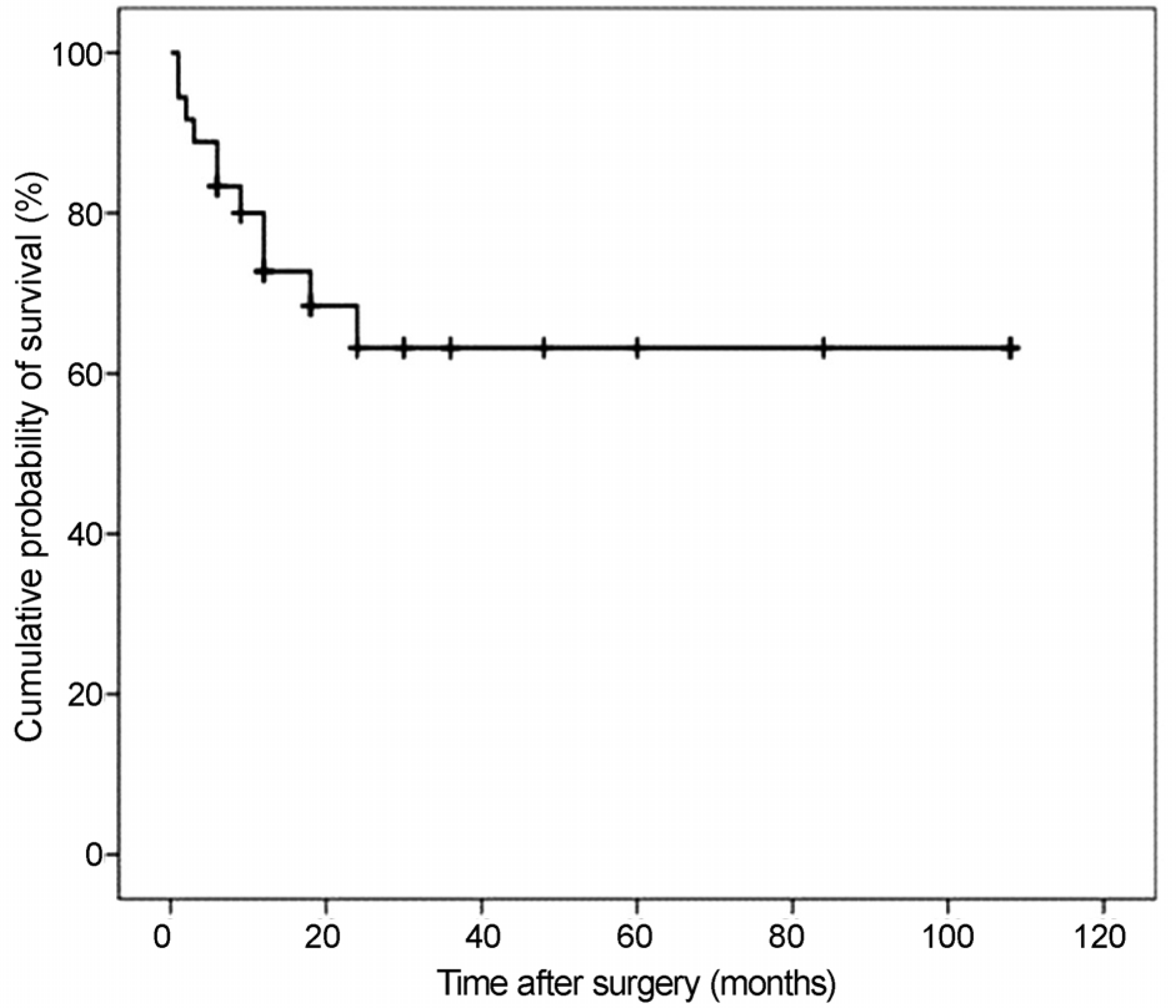
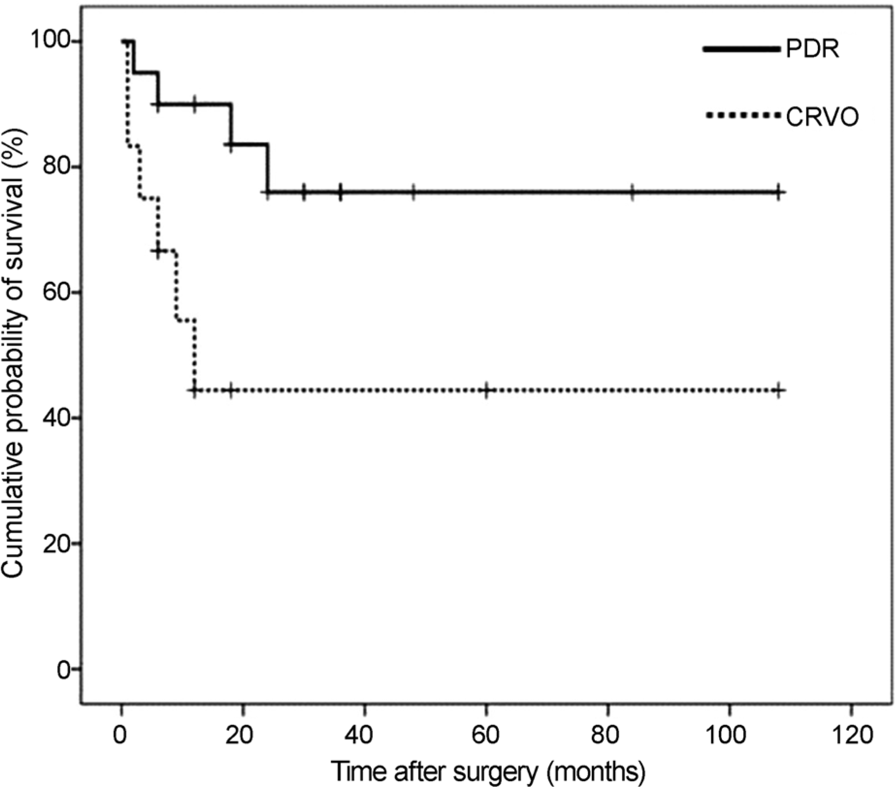
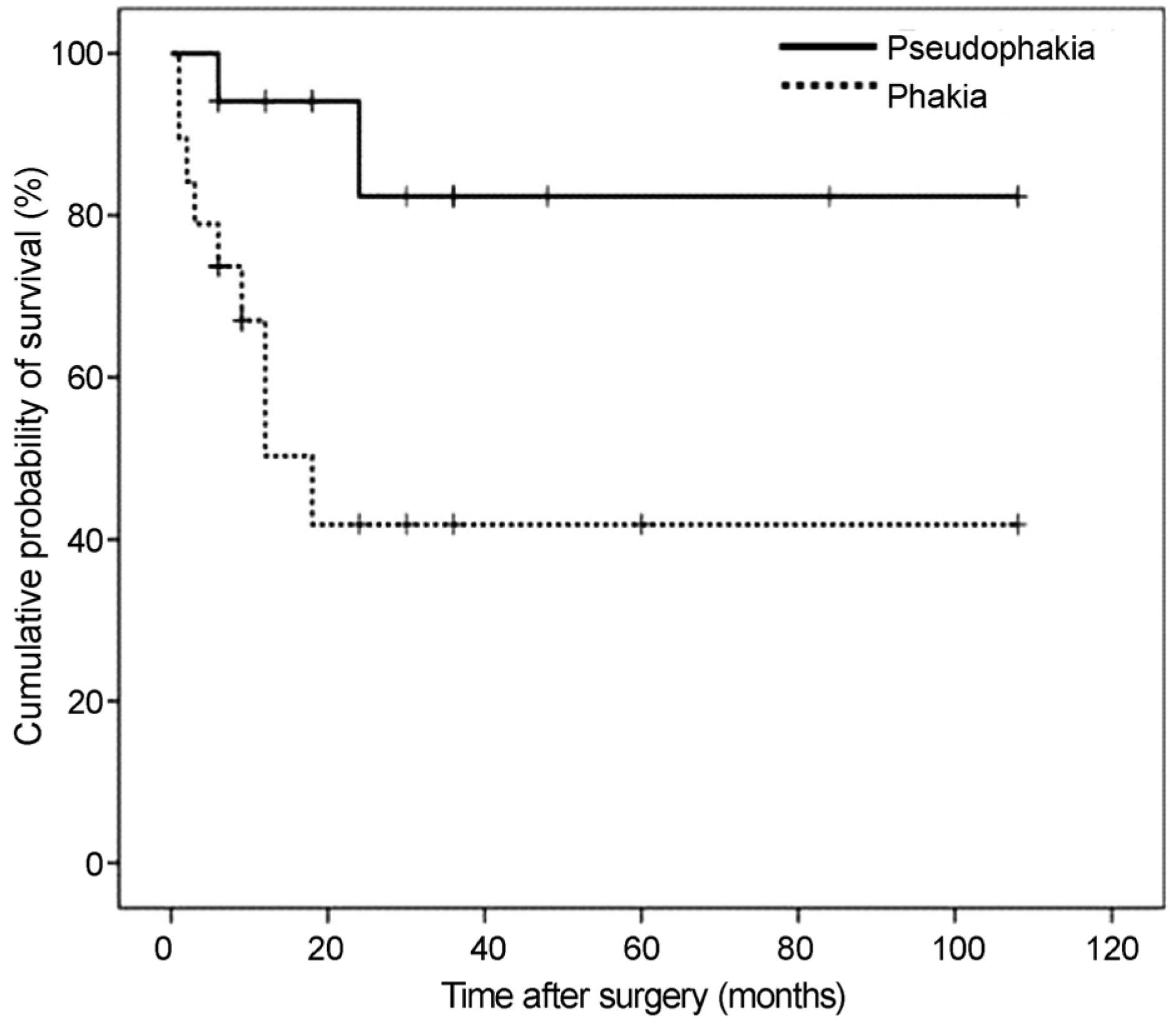
The overall success rate was 63.2% after a mean of 34.0 ± 31.7 months postoperative fdlow-up. The success rate was 83.3% at postoperative 6 months, 72.7% at postoperative 12 months and 63.2% at postoperative 3 years. The underlying retinal diseases were proliferative diabetic retinopathy (PDR; n = 20, 55.5%), central retinal vein occlusion (CRVO; n = 12, 33.3%), ocular ischemic syndrome (n = 2, 5.6%), and other retinal vascular diseases (n = 2, 5.6%). The survival times were significantly shorter in eyes with CRVO (20.2 ± 30.5 months) compared to PDR (33.1 ± 30.8 months), and in phakic eyes (33.1 ± 30.8 months) compared to pseudophakic eyes (37.7 ± 35.4 months) (p < 0.05). In the multivariate analysis, preoperative phakic eyes were significantly associated with a shorter survival time (hazard ratio = 5.626, p = 0.030).
Go to : 
References
1. Netland PA. The Ahmed glaucoma valve in neovascular glaucoma (An AOS Thesis). Trans Am Ophthalmol Soc. 2009; 107:325–42.
3. Liang CM. Chen YH. Lu DW, et al. New continuous air pumping technique to improve clinical outcomes of descemet-stripping automated endothelial keratoplasty in Asian patients with previous ahmed glaucoma valve implantation. PloS One. 2013; 8:e72089.

4. Netland PA. Ishida K. Boyle JW. The Ahmed glaucoma valve in patients with and without neovascular glaucoma. J Glaucoma. 2010; 19:581–6.

5. Lloyd MA. Heuer DK. Baerveldt G, et al. Combined molteno implantation and pars plana vitrectomy for neovascular glaucomas. Ophthalmology. 1991; 98:1401–5.

6. Moon DRC. Choi KS. Lee SJ. Ha SJ. Vitrectomy and Ahmed valve implantation in neovascular glaucoma patients with vitreous hemorrhage. J Korean Ophthalmol Soc. 2012; 53:801–6.

7. Scott IU. Alexandrakis G. Flynn HW Jr, et al. Combined pars plana vitrectomy and glaucoma drainage implant placement for refractory glaucoma. Am J Ophthalmol. 2000; 129:334–41.

8. Lee CM. Kim EA. Cho YW. Pars plana vitrectomy and Ahmed valve implantation for intractable glaucoma comorbid with retinal disorders. J Korean Ophthalmol Soc. 2011; 52:46–52.

9. Lee JJ. Park KH. Kim DM. Kim TW. Clinical outcomes of Ahmed glaucoma valve implantation using tube ligation and removable external stents. Korean J Ophthalmol. 2009; 23:86–92.

10. Shazly TA. Latina MA. Neovascular glaucoma: etiology, diagnosis and prognosis. Semin Ophthalmol. 2009; 24:113–21.

11. Coleman AL. Hill R. Wilson MR, et al. Initial clinical experience with the Ahmed Glaucoma Valve implant. Am J Ophthalmol. 1995; 120:23–31.

12. Susanna R Jr. Latin American Glaucoma Society Investigators. Partial Tenon’s capsule resection with adjunctive mitomycin C in Ahmed glaucoma valve implant surgery. Br J Ophthalmol. 2003; 87:994–8.
13. Sone H. Okuda Y. Kawakami Y, et al. Vascular endothelial growth factor level in aqueous humor of diabetic patients with rubeotic glaucoma is markedly elevated. Diabetes Care. 1996; 19:1306–7.

14. Stefánsson E. Physiology of vitreous surgery. Graefes Arch Clin Exp Ophthalmol. 2009; 247:147–63.

15. Barile GR. Chang S. Horowitz JD, et al. Neovascular complications associated with rubeosis iridis and peripheral retinal detachment after retinal detachment surgery. Am J Ophthalmol. 1998; 126:379–89.

16. Sivak-Callcott JA. O’Day DM. Gass JD. Tsai JC. Evidence-based recommendations for the diagnosis and treatment of neovascular glaucoma. Ophthalmology. 2001; 108:1767–76. quiz1777, 1800.
17. Fekrat S. Finkelstein D. Current concepts in the management of central retinal vein occlusion. Curr Opin Ophthalmol. 1997; 8:50–4.

18. Kang JY. Nam KY. Lee SJ. Lee SU. The effect of intravitreal bevacizumab injection before Ahmed valve implantation in patients with neovascular glaucoma. Int Ophthalmol. 2014; 34:793–9.

Go to : 
 | Figure 1.Kaplan-Meier survival curve following Ahmed valve implantation and 23 gauge vitrectomy. The success rates were 83.3% at 6 months and 72.7% at 12 months, 63.2% at 36 months, respectively. |
 | Figure 2.Kaplan-Meier survival curves according to the underlying retinal disease. The mean survival time was significantly different between central retinal vein occlusion (CRVO) (20.2 ± 30.5 months) and proliferative diabetic retinopathy (PDR) (33.1 ± 30.8 months, p = 0.028). |
 | Figure 3.Kaplan-Meier survival curves according to the preoperative lens status. The mean survival time was significantly different between preoperative phakic (34.2 ± 32.9 months) and pseudophakic eyes (18.8 ± 25.5 months, p = 0.011). |
Table 1.
Patient demographics
Table 2.
Data summary according to the underlying retinal disease
| PDR (n = 20) | CRVO (n = 12) | OIS (n = 2) | Eales disease (n = 1) | NF-1 related retinal vascular abnormality (n = 1) | |
|---|---|---|---|---|---|
| Age (years) | 57.1 ± 15.1 | 62.0 ± 16.3 | 66.5 ± 5.5 | 28 | 12 |
| Follow-up period (months) | 38.4 ± 32.1 | 34 ± 33.29 | 14 ± 7 | 10 | 12 |
| Phakic eye (n, %) | 7 (35.0) | 9 (75.0) | 1 (50.0) | 1 (100.0) | 1 (100.0) |
| Vitrectomized eyes (n, %) | 8 (40.0) | 1 (8.3) | 2 (100.0) | 1 (100.0) | 0 (0.0) |
| Preoperative PRP (n, %) | 17 (85.0) | 5 (41.6) | 2 (100.0) | 1 (100.0) | 0 (0.0) |
| Preoperative IVB (n, %) | 11 (55.0) | 6 (50.0) | 2 (100.0) | 1 (100.0) | 1 (100.0) |
| VA (log MAR) | |||||
| Preop | 2.46 ± 0.63 | 2.66 ± 0.31 | 2.65 ± 0.05 | 2.6 | 0.7 |
| Postop | 2.03 ± 0.96∗ | 2.69 ± 0.33 | 2.65 ± 0.05 | 1.22 | 2.7 |
| IOP (mm Hg) | |||||
| Preop | 35.7 ± 8.7 | 37 ± 13.6 | 32.5 ± 8.5 | 31 | 48 |
| Postop | 17.5 ± 4.1∗ | 20 ± 7.8∗ | 11.5 ± 5.5 | 18 | 13 |
| Number of medication | |||||
| Preop | 3.1 ± 0.7 | 3.0 ± 0.4 | 2 | 3 | 3 |
| Postop | 1.5 ± 1.2∗ | 0.9 ± 1.2∗ | 0 | 0 | 0 |
| Number of failure (n, %) | 5 (25.0) | 6 (50.0) | 1 (50.0) | 0 | 0 |
| Uncontrolled IOP | 2 | 3 | 1 | 0 | 0 |
| Changed to NLP | 3 | 3 | 0 | 0 | 0 |
PDR = proliferative diabetic retinopathy; CRVO = central retinal vein occlusion; OIS = ocular ischemic syndrome; NF-1 = neurofibromatosis type-1; PRP = panretinal photocoagulation; IVB = intravitreal bevacizumab injection; VA = visual acuity; Preop = preoperative; Postop = postoperative; IOP=intraocular pressure; NLP = no light perception.
Table 3.
Postoperative complications
Table 4.
Outcomes of Cox’s proportional hazard regression model
|
Univariate |
Multivariate∗ |
|||
|---|---|---|---|---|
| Risk ratio and 95% CI | p-value | Risk ratio and 95% CI | p-value | |
| Age (years) | 1.000 (0.967-1.034) | 0.996 | - | - |
| Gender | 1.233 (0.360-4.220) | 0.739 | - | - |
| DM | 0.401 (0.078-2.045) | 0.271 | - | - |
| HTN | 1.090 (0.282-4.220) | 0.901 | - | - |
| Preoperative PRP | 0.335 (0.095-1.175) | 0.088∗ | 0.749 (0.188-2.88) | 0.658 |
| Preoperative IVB | 0.710 (0.205-2.464) | 0.590 | - | - |
| Preoperative lens status | 5.626 (1.187-26.678) | 0.030∗ | 5.626 (1.187-26.678) | 0.030 |
| Underlying disease (CRVO) | 3.824 (1.052-13.892) | 0.042∗ | 2.198 (0.715-10.629) | 0.127 |
| Intraoperative IVB | 2.186 (0.560-8.523) | 0.260 | - | - |
| Preoperative vitrectomized eyes | 2.032 (0.429-9.615) | 0.371 | - | - |


 PDF
PDF ePub
ePub Citation
 Print
Print


 XML Download