Abstract
Purpose
To evaluate the incidence rate of postoperative glaucoma after pars plana vitrectomy (PPV) and to compare incidence rate of glaucoma between phakic and pseudophakic eyes.
Methods
Retrospective chart review of patients who underwent PPV between January 2007 and May 2014. The outcome meas-ure was the presence or absence of postoperative glaucoma, defined as definitive loss of neuro-retinal rim defect on fundus ex-am or showing glaucomatous change on a visual field test that warranted maintenance of ocular hypotensive therapy.
Results
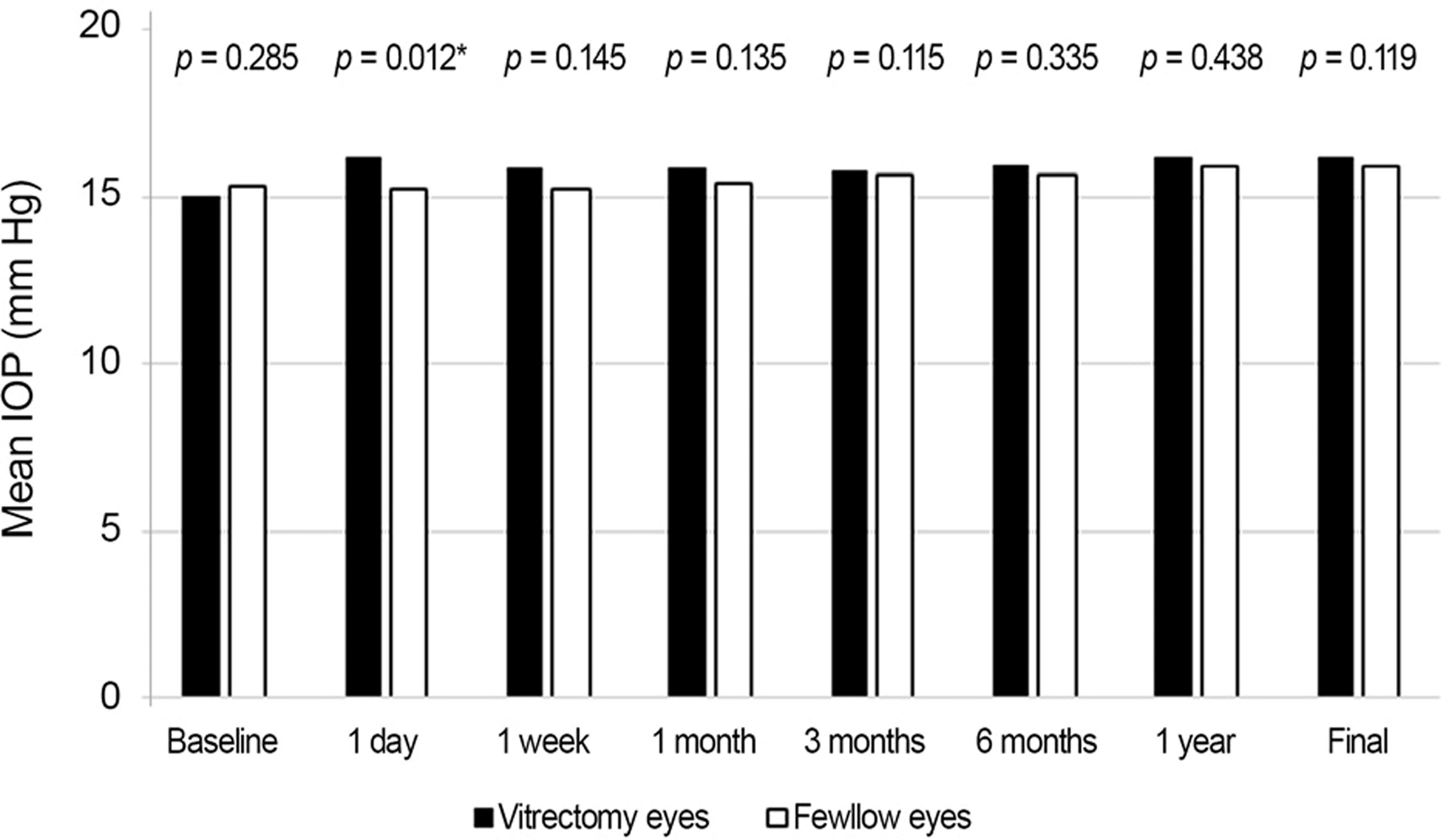
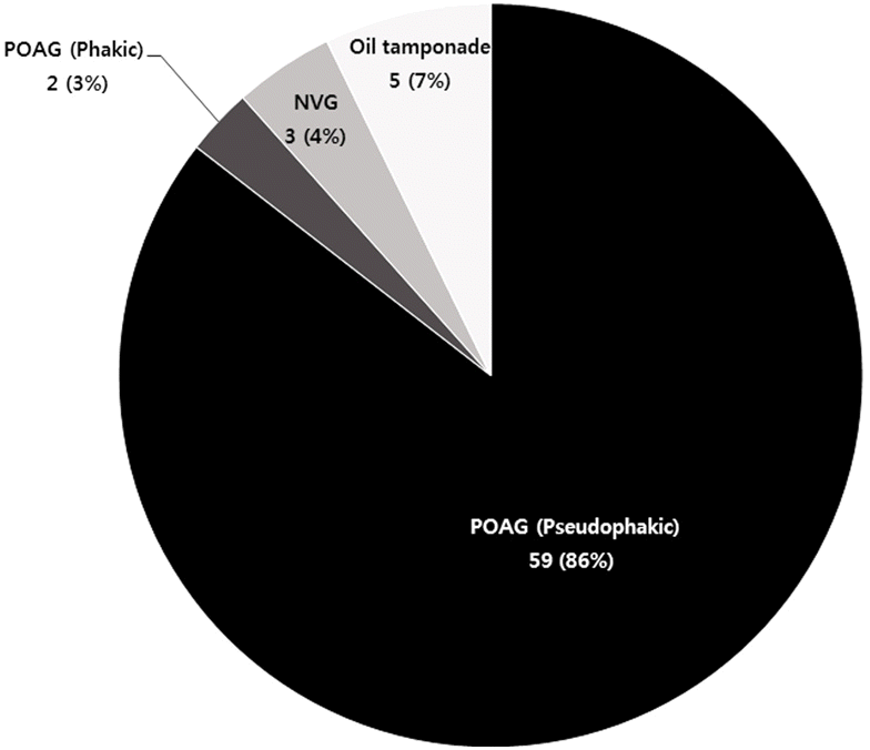
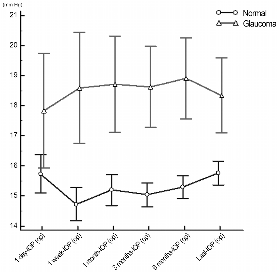
There were 333 patients average age 57.09 ± 13.43 included this study. Patients were followed for an average of 56.23 ± 10.85 months. There was no significant difference in mean intra ocular pressure (IOP) between the vitretomized eyes with un-operative eyes, except in mean IOP at one day postoperatively ( p = 0.012). In unoperative eyes, 10 of 315 (3.1%) were newly di-agnosed as postoperative glaucoma. For the vitrectomized eyes, 69 of the 351 (19.6%) were newly diagnosed as postoperative glaucoma. There was a significant difference in incidence rate of glaucoma between the two groups ( p < 0.001, chi-square test). There was a significantly higher IOP in glaucoma eyes compared with normal eyes ( p < 0.001, Repeated Measures Analysis of variance [RM-ANOVA]). A possible risk factor for the development of glaucoma after PPV was cataract surgery ( p = 0.0497, chi-square test).
Conclusions
The incidence of glaucoma in patients with PPV is higher than in normal eyes. The incidence seems to increase particularly in those who have a pesudophakic eye. Patients who underwent PPV, especially in the pseudophakic state, need to have their IOP monitored carefully and managed properly by an ophthalmologist.
Go to : 
References
2. Fujii GY, De Juan E Jr, Humayun MS. . Initial experience using the transconjunctival sutureless vitrectomy system for vitreoretinal surgery. Ophthalmology. 2002; 109:1814–20.
3. de Bustros S, Thompson JT, Michels RG. . Nuclear sclerosis after vitrectomy for idiopathic epiretinal membranes. Am J Ophthalmol. 1988; 105:160–4.

4. Melberg NS, Thomas MA. Nuclear sclerotic cataract after vi-trectomy in patients younger than 50 years of age. Ophthalmology. 1995; 102:1466–71.

5. Han DP, Lewis H, Lambrou FH Jr. . Mechanisms of intraocular pressure elevation after pars plana vitrectomy. Ophthalmology. 1989; 96:1357–62.
6. Tranos P, Asaria R, Alyward W. . Long term outcome of secon-dary glaucoma following vitreoretinal surgery. Br J Ophthalmol. 2004; 88:341–3.

7. Lalezary M, Kim SJ, Jiramongkolchai K. . Long-term trends in intraocular pressure after pars plana vitrectomy. Retina. 2011; 31:679–85.

8. Koreen L, Yoshida N, Escariao P. . Incidence of, risk factors for, and combined mechanism of late-onset open-angle glaucoma after vitrectomy. Retina. 2012; 32:160–7.

9. Chang S. LXII Edward Jackson lecture: open angle glaucoma after vitrectomy. Am J Ophthalmol. 2006; 141:1033–43.

10. Tham YC, Li X, Wong TY. . Global prevalence of glaucoma and projections of glaucoma burden through 2040: a systematic re-view and meta-analysis. Ophthalmology. 2014; 121:2081–90.
11. Wong EY, Keeffe JE, Rai JL. . Detection of undiagnosed glau-coma by eye health professionals. Ophthalmology. 2004; 111:1508–14.

12. Topouzis F, Coleman AL, Harris A. . Factors associated with undiagnosed open-angle glaucoma: the Thessaloniki Eye Study. Am J Ophthalmol. 2008; 145:327–35.

13. Musch DC, Gillespie BW, Lichter PR. . Visual field pro-gression in the Collaborative Initial Glaucoma Treatment Study the impact of treatment and other baseline factors. Ophthalmology. 2009; 116:200–7.
14. Wu L, Berrocal MH, Rodriguez FJ. . Intraocular pressure ele-vation after uncomplicated pars plana vitrectomy: results of the Pan American Collaborative Retina Study Group. Retina. 2014; 34:1985–9.
15. Benz MS, Escalona-Benz EM, Murray TG. . Immediate post-operative use of a topical agent to prevent intraocular pressure ele-vation after pars plana vitrectomy with gas tamponade. Arch Ophthalmol. 2004; 122:705–9.
16. Kim CS, Seong GJ, Lee NH. . Prevalence of primary open-an-gle glaucoma in central South Korea the Namil study. Ophthalmology. 2011; 118:1024–30.
17. Tranos P, Bhar G, Little B. Postoperative intraocular pressure spikes: the need to treat. Eye (Lond). 2004; 18:673–9.

18. Geijer C, Bill A. Effects of raised intraocular pressure on retinal, prelaminar, laminar, and retrolaminar optic nerve blood flow in monkeys. Invest Ophthalmol Vis Sci. 1979; 18:1030–42.
19. Sugiura Y, Okamoto F, Okamoto Y. . Ophthalmodynamometric pressure in eyes with proliferative diabetic retinopathy measured during pars plana vitrectomy. Am J Ophthalmol. 2011; 151:624–9.e1.

20. Jonas JB, Harder B. Ophthalmodynamometric differences between ischemic vs nonischemic retinal vein occlusion. Am J Ophthalmol. 2007; 143:112–6.

21. Saccà SC, Izzotti A, Rossi P, Traverso C. Glaucomatous outflow pathway and oxidative stress. Exp Eye Res. 2007; 84:389–99.

Go to : 
 | Figure 1.Mean IOP of the vitrectomized eyes compared with the unoperative fellow eyes at baseline and postoperatively. IOP = intra ocular pressure. * There is no significant differ-ence in mean IOP between the two groups, except in mean IOP at postoperative 1 day ( p = 0.012). |
 | Figure 2.Distribution of newly diagnosed glaucoma after PPV. PPV = pars plana vitrectomy; NVG = neovascular glau-coma; POAG = primary open-angle glaucoma. |
 | Figure 3.Postoperative mean IOP changes in normal and glau-coma group (RM-ANOVA by Greenhouse-Geisser method: between p < 0.001, within p = 0.167). Statistically significant higher IOP in glaucoma group than in normal group. IOP = in-tra ocular pressure; RM-ANOVA = repeated measures analysis of variance; op = operative. |
Table 1.
Baseline characteristics
| Vitrectomy eye (n = 351, 52.7%) | Unoperative eye (n = 315, 47.3%) | ) p-value* | |
|---|---|---|---|
| Age at the time of surgery (years) | 57.06 ± 13.43 | 54.24 ± 13.76 | 0.649 |
| Gender (male/female)† | 174/177 | 158/157 | 0.883 |
| Preoperative BCVA (decimal) | 0.21 ± 0.26 | 0.71 ± 0.33 | <0.001‡ |
| Preoperative IOP (mm Hg) | 15.03 ± 4.13 | 15.31 ± 3.25 | 0.285 |
Table 2.
Comparison of incidence rate of glaucoma after vi-trectomy among vitreoretinal disorder
| Type of disorder | Incidence rate (incidence/total, %) |
|---|---|
| RRD | 27/140 (19%) |
| PDR | 27/99 (27%) |
| ERM RVO | 7/75 (9%) 3/12 (25%) |
| MH | 2/10 (20%) |
| PVR | 1/7 (14%) |
Table 3.
Risk factors for the development of glaucoma after PPV
| Patient | Normal (n = 282) | Glaucoma (n = 69) | p-value* | Odd ratio | 95% CI |
|---|---|---|---|---|---|
| Age at the time of surgery (years) | 57.64 ± 12.90 | 54.84 ± 15.36 | 0.1202 | ||
| Gender (male/female, n)† | 133/149 | 41/28 | 0.0908 | 1.6404 | 0.9613-2.7992 |
| Preoperative BCVA (decimal) | 0.22 ± 0.27 | 0.17 ± 0.25 | 0.0957 | ||
| Preoperative IOP (mm Hg) | 14.72 ± 4.01 | 15.30 ± 4.59 | 0.2963 | ||
| ILM peeling (Y/N) | 73/209 | 7/62 | 0.0523 | 0.323 | 0.2083-1.2680 |
| Presence of crystalline lens† | 0.0497* | 4.2880‡ | 1.0020-18.3500 | ||
| Pseudophakia | 254 | 67 | |||
| Phakia | 28 | 2 | |||
| Gauge of trocar (20 G/23 G)† | 76/206 | 22/47 | 0.5034 | 1.2688 | 0.7171-2.2448 |


 PDF
PDF ePub
ePub Citation
 Print
Print


 XML Download