

 PDF
PDF ePub
ePub Citation
 Print
Print


Abstract
Purpose
To compare the measurement results of 3 ocular biometry devices, A-scan ultrasound and two types of partial coherence interferometers in normal and cataractous eyes.
Methods
This study included 42 normal eyes and 40 cataractous eye. Axial length and anterior chamber were measured using three ocular biometry measurements, ultrasonography (HiScan®, Optikon 2000, Rome, Italy), IOL Master® (Carl Zeiss, Jena, Germany), and AL-scan® (Nidek, Gamagori, Japan), and mean corneal curvature and corneal diameter were measured using two partial coherence interferometers. The results were compared in each group.
Results
Significant differences in measurements existed among the 3 ocular biometry devices (A-scan ultrasound, IOL Master® and AL-scan®) in normal eyes (p < 0.001) and cataractous eyes (p = 0.034). However, the measurements were not significantly different between the 2 partial coherence interferometers (IOL Master® and AL-scan®) in both groups. We confirmed lower agreement among the 3 ocular biometry devices in cataractous eyes compared with normal eyes in terms of a larger range of 95% agreement and error in cataractous eyes.
Conclusions
Significant differences in measurements were observed when using the 3 ocular biometry devices in both normal and cataractous eyes. Because of low agreements between ocular biometry devices in cataractous eyes, complementing the measurements between ocular biometry devices is necessary when measuring cataractous eyes.
Go to : 
References
1. Olsen T. Sources of error in intraocular lens power calculation. J Cataract Refract Surg. 1992; 18:125–9.

2. Santodomingo-Rubido J. Mallen EA. Gilmartin B. Wolffsohn JS. A new non-contact optical device for ocular biometry. Br J Ophthalmol. 2002; 86:458–62.
3. Kim JW. Lee H. Jung JW, et al. Comparison of ocular biometry using low-coherence reflectometry with other devices for intraocular lens power calculation. J Korean Ophthalmol Soc. 2015; 56:1558–65.
4. Drexler W. Hitzenberger CK. Baumgartner A, et al. Investigation of dispersion effects in ocular media by multiple wavelength partial coherence interferometry. Exp Eye Res. 1998; 66:25–33.
5. Drexler W. Findl O. Menapace R, et al. Partial coherence interferometry: a novel approach to biometry in cataract surgery. Am J Ophthalmol. 1998; 126:524–34.
6. Holzer MP. Mamusa M. Auffarth GU. Accuracy of a new partial coherence interferometry analyser for biometric measurements. Br J Ophthalmol. 2009; 93:807–10.
7. Hitzenberger CK. Optical measurement of the axial eye length by laser Doppler interferometry. Invest Ophthalmol Vis Sci. 1991; 32:616–24.
8. Kwag JY. Choi SH. Comparison of ocular biometry measured by ultrasound and two kinds of partial coherence interferometers. J Korean Ophthalmol Soc. 2011; 52:169–74.
9. Kim SI. Kang SJ. Oh TH, et al. Accuracy of ocular biometry and postoperative refraction in cataract patients with AL-scan(R). J Korean Ophthalmol Soc. 2013; 54:1688–93.
10. Haigis W. Lege B. Miller N. Schneider B. Comparison of immersion ultrasound biometry and partial coherence interferometry for intraocular lens calculation according to Haigis. Graefes Arch Clin Exp Ophthalmol. 2000; 238:765–73.
11. Hoffer KJ. Shammas HJ. Savini G. Comparison of 2 laser instruments for measuring axial length. J Cataract Refract Surg. 2010; 36:644–8.
12. Huang J. Savini G. Li J, et al. Evaluation of a new optical biometry device for measurements of ocular components and its comparison with IOLMaster. Br J Ophthalmol. 2014; 98:1277–81.
13. Lackner B. Schmidinger G. Skorpik C. Validity and repeatability of anterior chamber depth measurements with Pentacam and Orbscan. Optom Vis Sci. 2005; 82:858–61.
14. Utine CA. Altin F. Cakir H. Perente I. Comparison of anterior chamber depth measurements taken with the Pentacam, Orbscan IIz and IOLMaster in myopic and emmetropic eyes. Acta Ophthalmol. 2009; 87:386–91.
15. Hwang JS. Lee JH. Comparison of the IOL Master(R) and A-scan ultrasound: refractive results of 96 consecutive cases. J Korean Ophthalmol Soc. 2007; 48:27–32.
16. Chung JK. Choe CM. You YS. Lee SJ. Biometry with partial coherence interferometry and ultrasonography in high myopes. J Korean Ophthalmol Soc. 2006; 47:355–61.
17. Whang WJ. Byun YS. Joo CK. Comparison of refractive outcomes using five devices for the assessment of preoperative corneal power. Clin Experiment Ophthalmol. 2012; 40:425–32.
18. Eibschitz-Tsimhoni M. Tsimhoni O. Archer SM. Del Monte MA. Effect of axial length and keratometry measurement error on intraocular lens implant power prediction formulas in pediatric patients. J AAPOS. 2008; 12:173–6.
19. Huang J. McAlinden C. Su B, et al. The effect of cycloplegia on the lenstar and the IOLMaster biometry. Optom Vis Sci. 2012; 89:1691–6.
20. Kaswin G. Rousseau A. Mgarrech M, et al. Biometry and intraocular lens power calculation results with a new optical biometry device: comparison with the gold standard. J Cataract Refract Surg. 2014; 40:593–600.
Go to :
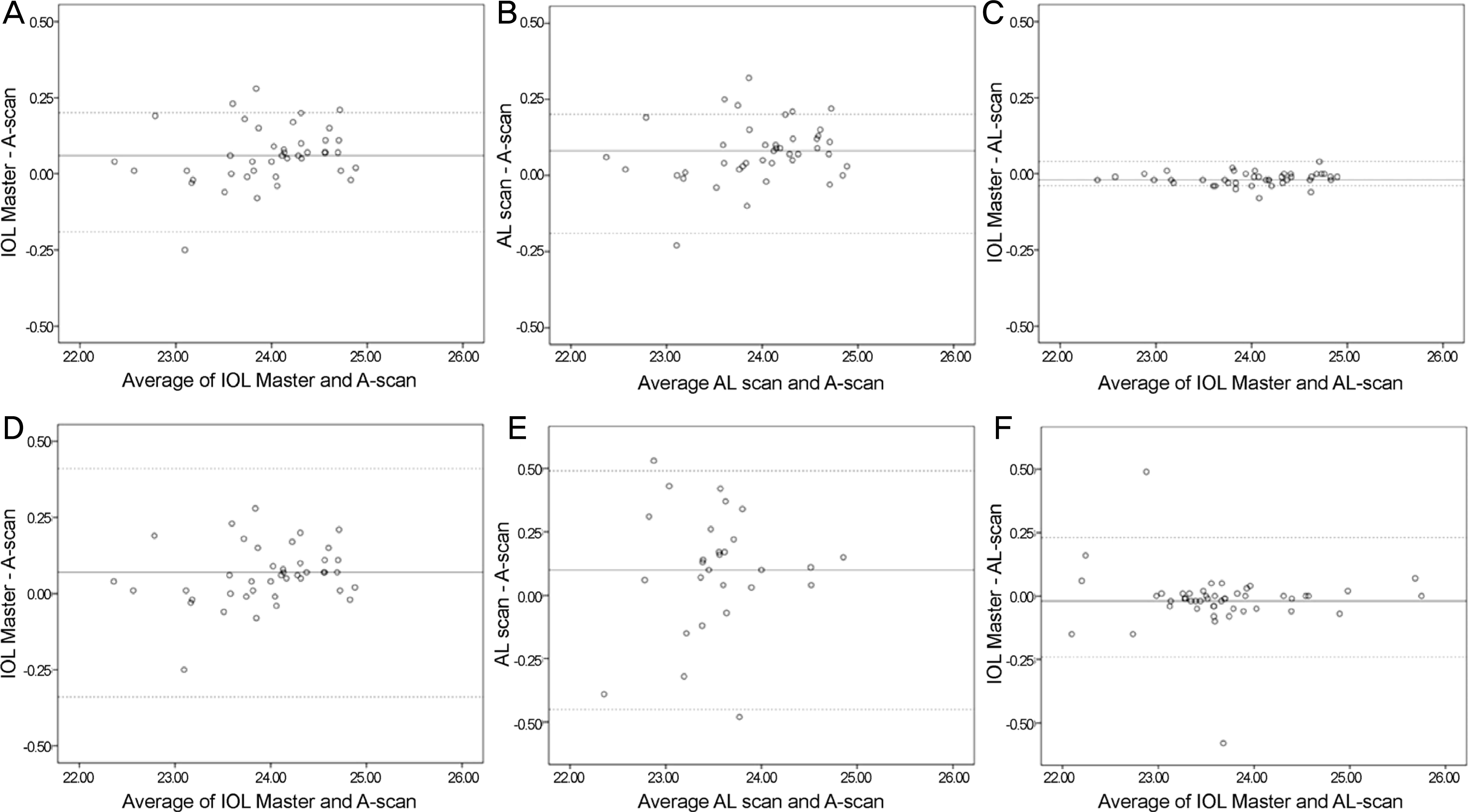 | Figure 1.Bland-Altman plots of axial length (mm) showing the agreement among the three devices in normal and cataractous eyes. (A) IOL Master® and A-scan in normal eyes. (B) AL scan and A-scan in normal eyes. (C) IOL Master® and AL scan in normal eyes. (D) IOL Master® and A-scan in cataractous eyes. (E) AL scan and A-scan in cataractous eyes. (F) IOL Master® and AL scan in cataractous eyes showing the 95% limits of agreement for axial length difference: -0.19 ~ 0.20 mm, -0.19 ~ 0.20 mm, -0.04 ~ 0.04 mm, -0.40 ~ 0.43 mm, -0.45 ~ 0.49 mm and -0.24 ~ 0.23 mm, respectively. |
Table 1.
Characteristics of normal and cataractous eyes
| Normal eyes | Cataractous eyes | p-value‡ | |
|---|---|---|---|
| No. of eyes | 42 | 40 | |
| Sex (male:female) | 28:14 | 23:17 | |
| Age (years) | 36.9 ± 5.6 (21-70) | 59.3 ± 10.9 (17-89) | |
| BCVA (decimal) | 1.0 ± 0.0 | 0.4 ± 0.25 | |
| Spherical equivalent (D)∗ | -0.27 ± 0.44 (-2.12~1.25) | 0.15 ± 0.47 (-2.50~1.37) | 0.262 |
| Mean keratometric value (D)∗ | 44.05 ± 1.24 (41.13~46.62) | 44.00 ± 1.46 (41.12~46.50) | 0.121 |
| Axial length (mm)† | 23.93 ± 0.61 (22.34~24.87) | 23.41 ± 0.55 (22.38~24.78) | 0.052 |
Table 2.
Comparison of biometric measurements when using the 3 biometry devices in normal and cataractous eyes
| A-scan ultrasound biometry | IOL Master® | AL-scan® | p-value∗ |
p-value† |
|||
|---|---|---|---|---|---|---|---|
| IOL Master® and A-scan | AL-scan® and A-scan | IOL Master® and AL-scan® | |||||
| Normal eyes | |||||||
| AL (mm) | 23.93 ± 0.61 | 23.99 ± 0.63 | 24.01 ± 0.63 | <0.001 | <0.001 | <0.001 | 0.059 |
| ACD (mm) | 3.32 ± 0.38 | 3.29 ± 0.31 | 3.39 ± 0.37 | 0.051 | 0.105 | 0.141 | 0.842 |
| Km (D) | 44.05 ± 1.24‡ | 43.84 ± 1.26 | 43.97 ± 1.27 | 0.284 | 0.276 | 0.279 | 0.686 |
| WTW (mm) | 12.13 ± 0.48 | 11.78 ± 0.35 | <0.001 | ||||
| Cataractous eyes | |||||||
| AL (mm) | 23.41 ± 0.55 | 23.63 ± 0.72 | 23.68 ± 0.75 | 0.034 | 0.049 | 0.034 | 0.393 |
| ACD (mm) | 3.34 ± 0.21 | 3.45 ± 0.34 | 3.60 ± 0.48 | 0.015 | 0.506 | 0.027 | 0.003 |
| Km (D) | 44.05 ± 1.24‡ | 43.84 ± 1.26 | 43.97 ± 1.27 | 0.163 | 0.124 | 0.635 | 0.051 |
| WTW (mm) | 12.01 ± 0.64 | 11.57 ± 0.30 | 0.002 | ||||
Table 3.
Comparison of prediction error among the 3 biometry devices
| A-scan ultrasound biometry | IOL Master® | AL-scan® | p-value∗ | |
|---|---|---|---|---|
| Mean PE | 0.13 ± 0.53 | -0.14 ± 0.50 | 0.00 ± 0.60 | 0.505 |
| Absolute PE | 0.37 ± 0.39 | 0.41 ± 0.39 | 0.45 ± 0.36 | 0.844 |
 XML Download