Abstract
Purpose
To compare the short-term clinical outcomes of transscleral fixation intraocular lens (IOL) with two haptics or one haptic.
Methods
We retrospectively reviewed the medical records of 26 patients with transscleral fixation of IOL (11 with one-haptic fixation, 15 with two-haptic fixation) except in patients whose visual acuity is not expected to improve due to retinal problems or ocular trauma. We analyzed the manifest refraction, visual acuity, refractive error preoperatively and postoperatively, preoperative IOL decentration, operating time, and postoperative complications.
Results
When comparing the two groups preoperatively, the mean lens decentration in the one-haptic group was 2.73 ± 2.88 mm and 4.59 ± 2.18 mm in the two-haptics group. The decentration in the two-haptic group was greater than in the one-haptic group, but not significantly. Visual acuity and refractive power were not significantly different between the groups. The mean operation time (minutes) was 65.00 ± 22.69 and 93.66 ± 29.54 in the one-haptic and two-haptic groups, respectively. The operation time in the one-haptic group was significantly shorter than in the two-haptic group (p = 0.020). When comparing visual acuity preoperatively and postoperatively, both groups showed significant improvement (p < 0.01). However, refractive error and postoperative IOL decentration were similar between the groups.
Go to : 
References
1. Jakobsson G. Zetterberg M. Lundström M, et al. Late dislocation of in-the-bag and out-of-the bag intraocular lenses: ocular and surgical characteristics and time to lens repositioning. J Cataract Refract Surg. 2010; 36:1637–44.

2. Hu BV. Shin DH. Gibbs KA. Hong YJ. Implantation of posterior chamber lens in the absence of capsular and zonular support. Arch Ophthalmol. 1988; 106:416–20.

3. Stark WJ. Gottsch JD. Goodman DF, et al. Posterior chamber intraocular lens implantation in the absence of capsular support. Arch Ophthalmol. 1989; 107:1078–83.

4. Duffey RJ. Holland EJ. Agapitos PJ. Lindstrom RL. Anatomic study of transsclerally sutured intraocular lens implantation. Am J Ophthalmol. 1989; 108:300–9.

5. Yang JY. Chu YK. Modified surgical technique for transscleral fixation of a single-piece acrylic intraocular lens in the absence of capsular support. J Korean Ophthalmol Soc. 2012; 53:1794–800.

6. Kim SJ. Lee SJ. Park CH, et al. Long-term stability and visual outcomes of a single-piece, foldable, acrylic intraocular lens for scleral fixation. Retina. 2009; 29:91–7.

7. Ryu EH. Lee JH. LEE SY. Surgical results of transscleral fixation of foldable acrylic intraocular lens with unique shape of haptic. J Korean Ophthalmol Soc. 2008; 49:1071–7.
8. Liu S. Cheng S. Modified method of sutureless intrascleral posterior chamber intraocular lens fixation without capsular support. Eur J Ophthalmol. 2013; 23:732–7.

9. Kongsap P. Aknotless, one-haptic fixation of posterior chamber intraocular lenses: one-year results. Int J Ophthalmol. 2015; 8:104–6.
10. Moawad AI. Ghanem AA. One-haptic fixation of posterior chamber intraocular lenses without scleral flaps. J Ophthalmol. 2012; 2012:8918–39.

11. Verbruggen KH. Rozema JJ. Gobin L, et al. Intraocular lens centration and visual outcomes after bag-in-the-lens implantation. J Cataract Refract Surg. 2007; 33:1267–72.

12. Montés-Micó R. Cerviño A. Ferrer-Blasco T. Intraocualr lens centration and stability: efficacy of current technique and technology. Curr Opin Ophthalmol. 2009; 20:33–6.
13. Malbran ES. Malbrane E Jr. Negri I. Lens guide suture for transport and fixation in secondary IOL implantation after intracapsular extraction. Int Ophthalmol. 1986; 9:151–60.

14. Moon AR. Moon NJ. Choi KY. Long-term results and complications using scleral-fixated posterior chamber intraocular lenses. J Korean Ophthalmol Soc. 1996; 37:1283–92.
15. Park YJ. Joo CK. Comparison of clinical results between sulcus insertion and transscleral fixation of intraocular lenses. J Korean Ophthalmol Soc. 1999; 40:1535–43.
16. Steiner A. Steinhorst UH. Steiner M, et al. Ultrasound biomicroscopy for localization of artificial lens haptics after trans-scleral suture fixation. Ophthalmologe. 1997; 94:41–4.
17. Han F. Liu W. Shu X, et al. Evaluation of pars plana sclera fixation of posterior chamber intraocular lens. Indian J Ophthalmol. 2014; 62:688–91.

18. Mello MO Jr. Scott IU. Smiddy WE, et al. Surgical management and outcomes of dislocated intraocular lenses. Ophthalmology. 2000; 107:62–7.

19. Ahn YS. Park YL. Kim HS. Refractive change after transscleral fixation of intraocular lens. J Korean Ophthalmol Soc. 2015; 56:548–58.

20. Hayashi K. Hayashi H. Nakao F. Hayashi F. Intraocular lens tilt and decentration, anterior chamber depth, and refractive error after trans-scleral suture fixation surgery. Ophthalmology. 1999; 106:878–82.
Go to : 
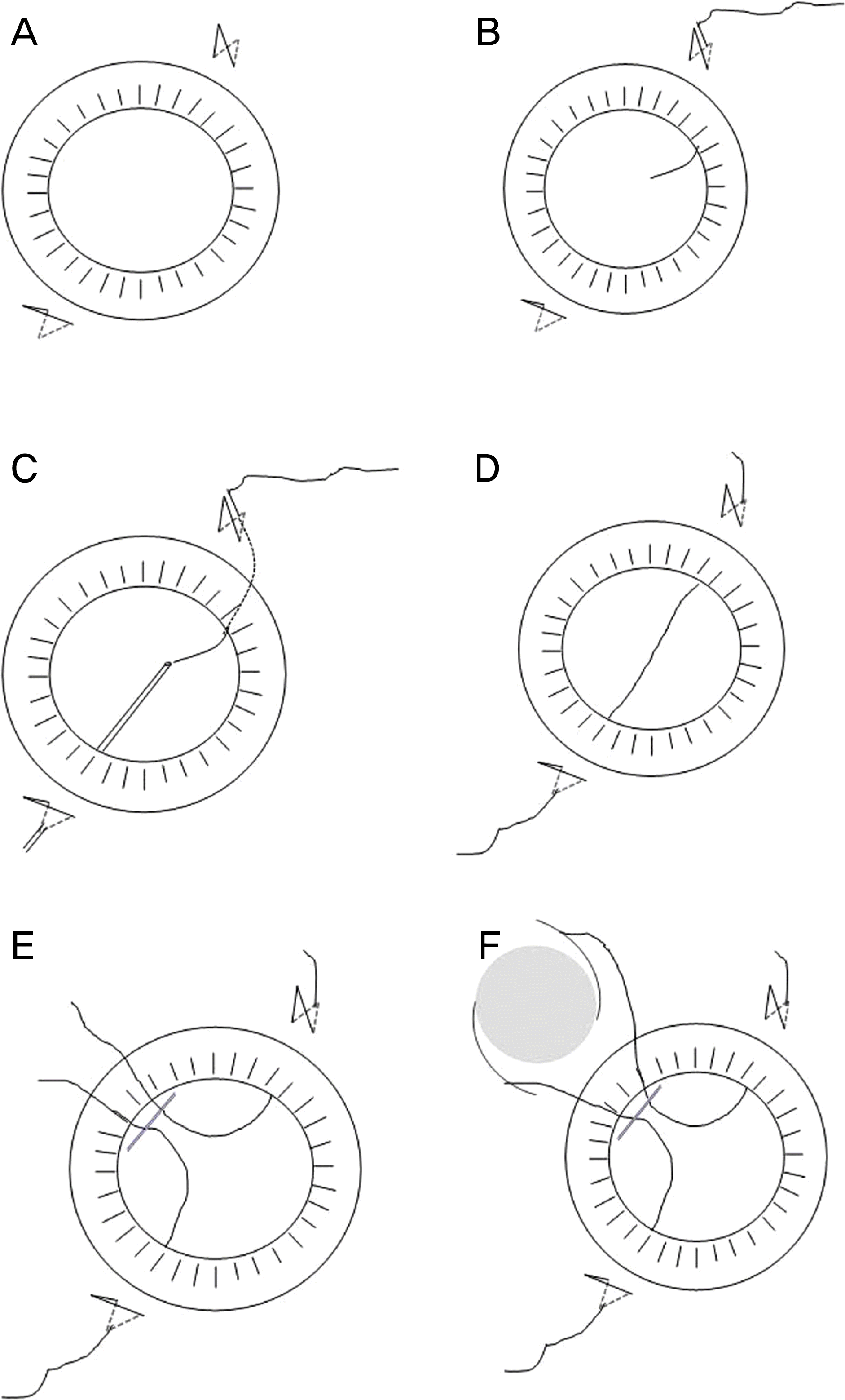
 | Figure 1.Steps of two-haptic transscleral fixation of intraocular lens (IOL). (A) Scleral flap is created at two sites. (B) A long curved double-armed 10-0 polypropylene needle is passed through the scleral flap site. (C) A 25-gauge syringe is passed from the opposite site and docked the curved needle. (D) The docked 25-gauge syringe and curved needle are moved from the scleral flap. (E) A 3.0 mm corneal incision is created at the temporal side and the 10-0 polypropylene is pulled through the corneal incision site with a Sinskey hook and is cut. (F) The 10-0 polypropylene is tied on both haptics of the IOL. |
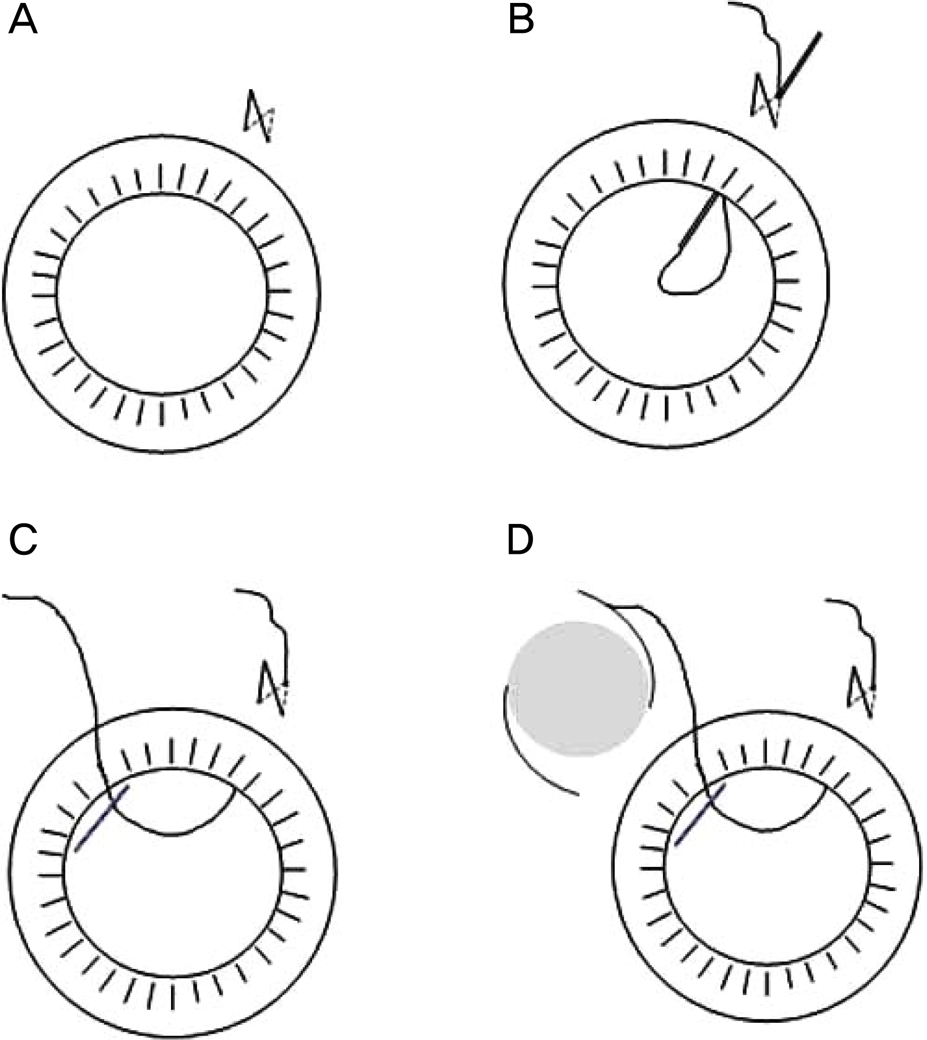
 | Figure 2.Steps of one haptic transscleral fixation of intraocular lens (IOL). (A) Scleral flap is created at one site. (B) Double-armed 10-0 polypropylene needle is cut in half, pushed through the end of the 25-gauge hollow syringe tip and the syringe is placed through the scleral flap site. (C) A 3.0 mm corneal incision is created at the temporal side and the 10-0 polypropylene is pulled through the corneal incision site. (D) The 10-0 polypropylene is tied on a one side of the haptic of the IOL. |
Table 1.
Preoperative clinical characteristics of one-haptic transscleral fixation of the intraocular lens (IOL) group
Table 2.
Preoperative clinical characteristics of two-haptic transscleral fixation of the intraocular lens (IOL) group
Table 3.
Comparison of decentration, target diopter and operation time between the two groups
| Surgical techniques | Group A (%, n = 11) | Group B (%, n = 15) | p-value∗ |
|---|---|---|---|
| Preop. IOL decentration (mm) | 2.73 ± 2.82 | 4.59 ± 2.18 | 0.109 |
| POD 3 months IOL decentration (mm) | 0.50 ± 1.01 | 0.24 ± 0.38 | 0.574 |
| Operating time (minutes) | 65.00 ± 22.69 | 93.66 ± 29.54 | 0.020 |
Table 4.
Comparison of the postoperative clinical outcome between the two groups
| Group A | Group B | p-value∗ | |
|---|---|---|---|
| UCVA (log MAR) | |||
| Preop. | 1.19 ± 0.74 | 0.98 ± 0.68 | 0.330 |
| POD 1 day | 0.60 ± 0.29† | 0.73 ± 0.47 | 0.646 |
| POD 1 month | 0.43 ± 0.25† | 0.52 ± 0.44† | 0.959 |
| POD 3 months | 0.36 ± 0.28† | 0.38 ± 0.31† | 0.959 |
| BCVA (log MAR) | |||
| Preop. | 0.49 ± 0.64 | 0.31 ± 0.32 | 0.413 |
| POD 1 day | 0.32 ± 0.23 | 0.47 ± 0.41 | 0.413 |
| POD 1 month | 0.15 ± 0.13† | 0.25 ± 0.40 | 0.799 |
| POD 3 months | 0.12 ± 0.13† | 0.12 ± 0.19† | 0.760 |
| Mean spherical (diopter) | |||
| Preop. | 3.25 ± 6.48 | 6.48 ± 6.28 | 0.330 |
| POD 1 day | -0.15 ± 1.26 | 0.15 ± 1.70† | 0.799 |
| POD 1 month | -0.50 ± 1.40 | -0.15 ± 1.81† | 0.919 |
| POD 3 months | -0.50 ± 1.37 | -0.11 ± 1.28† | 0.610 |
| Refractive error (diopter) | |||
| POD 1 day | 0.15 ± 0.83 | 0.51 ± 1.26 | 0.574 |
| POD 1 month | -0.22 ± 1.75 | 0.21 ± 1.39 | 0.443 |
| POD 3 months | -0.18 ± 0.84 | 0.25 ± 0.86 | 0.164 |
Values are presented as mean ± SD unless otherwise indicated. ‘Group A’ is ‘one-haptic transscleral fixation of IOL (count hapic sulcus fixation)’ and ‘Group B’ is ‘two-haptics transscleral fixation of IOL’.
Table 5.
Surgical techniques of one-haptic transscleral fixation of intraocular lens (IOL) group
Table 6.
Surgical techniques of two-haptic transscleral fixation of the intraocular lens (IOL) group


 PDF
PDF ePub
ePub Citation
 Print
Print


 XML Download