Abstract
Purpose
To report a case of double-layered conjunctival autograft and amniotic membrane transplantation for the effective treatment of esotropia and hypotropia after removal of the recurrent pterygium.
Case summary
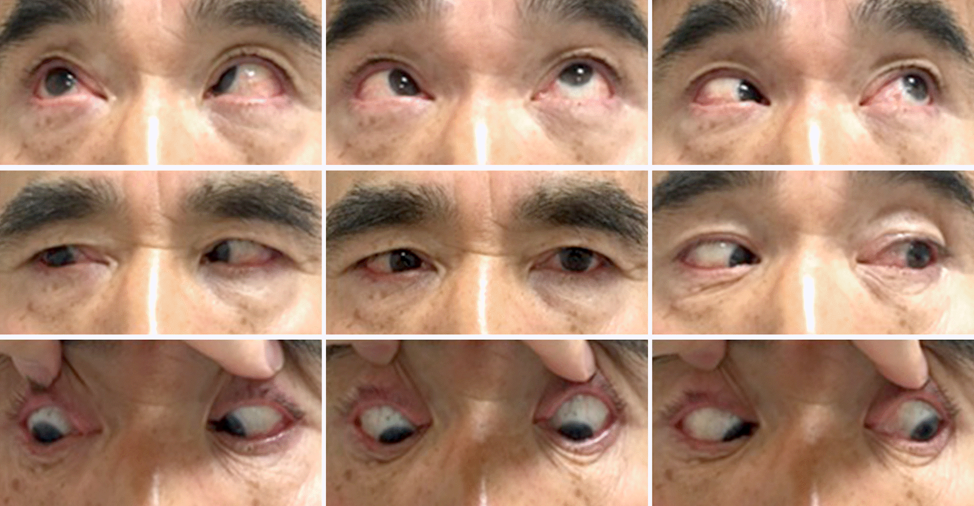
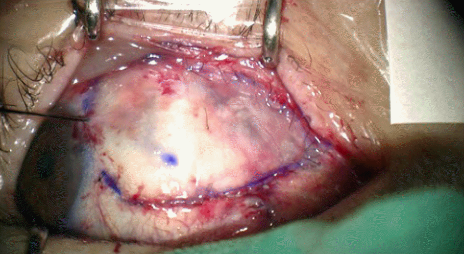
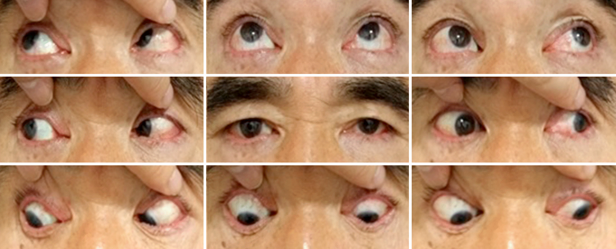
A 58-year-old male who had pterygium surgery of the right eye twice presented with diplopia on right gaze for 3 months. At the first visit, he had orthotropia in the primary position and right esotropia of 12 prism diopters (PD) on right gaze with limited abduction of −1 in the right eye. Fourteen months later, deviation was aggravated by esotropia of 30 PD and 12 PD of right hypotropia in the primary position at distance, and esotropia of 35 PD and 12 PD of right hypotropia at near with limited abduction of −2 and supraduction of −3 in the right eye. The patient complained of diplopia at all gazes and demonstrated chin-up posture. The conjunctival edge was recessed near the medial canthus and fornix, preventing conjunctival autograft after removal of subconjunctival scar tissue. Thus, 5 mm right medial rectus recession and additional half-sized conjunctival autograft were performed after amniotic membrane transplantation. The patient than showed no diplopia and orthotropia at both distance and near, with limited adduction of −1 in the right eye. He experienced no recurrence during 7 months of follow-up.
Conclusions
To prevent poor epithelial regeneration and dehiscence of graft in the patients with severe restrictive strabismus and very extensive conjunctival defect, double-layered conjunctival autograft and amniotic membrane transplantation may be effective for the treatment of severe esotropia and hypotropia.
Go to : 
References
1. Ela-Dalman N, Velez FG, Rosenbaum AL. Incomitant esotropia following pterygium excision surgery. Arch Ophthalmol. 2007; 125:369–73.

2. Jenkins PF, Stavis MI, Jenkins DE. 3rd. Esotropia following pterygium surgery. Binocul Vis Strabismus Q. 2002; 17:227–8.
3. Raab EL, Metz HS, Ellis FD. Medial rectus injury after pterygium excision. Arch Ophthalmol. 1989; 107:1428.

4. Katircioğlu YA, Altiparmak UE, Duman S. Comparison of three methods for the treatment of pterygium: amniotic membrane graft, conjunctival autograft and conjunctival autograft plus mitomycin C. Orbit. 2007; 26:5–13.

5. Kenyon KR, Wagoner MD, Hettinger ME. Conjunctival autograft transplantation for advanced and recurrent pterygium. Ophthalmology. 1985; 92:1461–70.

6. Dake CL, Crone RA, de Keizer RJ. Treatment of (recurrent) pterygium oculi by lamellar keratoplasty. Doc Ophthalmol. 1980; 48:223–30.

7. Jo SH, Lee JE, Choi HY, Jung JH. Clinical characteristics and treatment of esotropia rollowing bare sclera pterygium surgery. J Korean Ophthalmol Soc. 2013; 54:771–6.
8. Allan BD, Short P, Crawford GJ, et al. Pterygium excision with conjunctival autografting: an effective and safe technique. Br J Ophthalmol. 1993; 77:698–701.

9. Oh TH, Choi KY, Yoon BJ. The effect of conjunctivial autograft for recurrent pterygium. J Korean Ophthalmol Soc. 1994; 35:1335–9.
10. Shimazaki J, Shinozaki N, Tsubota K. Transplantation of amniotic membrane and limbal autograft for patients with recurrent pterygium associated with symblepharon. Br J Ophthalmol. 1998; 82:235–40.

11. Katrcoglu YA, Altiparmak U, Engur Goktas S, et al. Comparison of two techniques for the treatment of recurrent pterygium: amniotic membrane vs conjunctival autograft combined with mitomycin C. Semin Ophthalmol. 2015; 30:321–7.
12. Mamede AC, Carvalho MJ, Abrantes AM, et al. Amniotic membrane: from structure and functions to clinical applications. Cell Tissue Res. 2012; 349:447–58.

13. Benson-Martin J, Zammaretti P, Bilic G, et al. The Young's modulus of fetalpreterm and term amniotic membranes. Eur J Obstet Gynecol Reprod Biol. 2006; 128:103–7.
14. Zhu X, Beuerman RW, Chan-Park MB, et al. Enhancement of the mechanical and biological properties of a biomembrane for tissue engineering the ocular surface. Ann Acad Med Singapore. 2006; 35:210–4.
Go to : 
 | Figure 1.9 cardinals photo of the patient at first visit. The patient showed 30 PD esotropia and 12 PD hypotropia at distance and limited abduction (-2) and supraduction (-3) in the right eye. |
 | Figure 2.Right after double-layered conjunctival autograft and amniotic membrane transplantation. A 1.7×1.5 cm-sized defect was reconstructed by amniotic membrane transplantation with medial half conjunctival autograft after medial rectus recession of 5.0 mm in the right eye. |
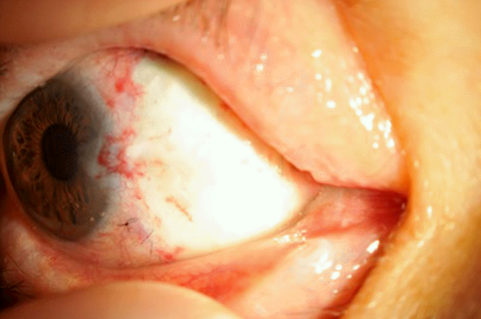
 | Figure 3.One month after double-layered conjunctival autograft and amniotic membrane transplantation. One month after surgery, the graft is located in its place and very clear. |
 | Figure 4.9 cardinals photo of the patient 7 months after surgery. Orthotropia at distance and near with only slight adduction limitation (-1) is noted in the right eye. Diplopia at all gaze positions, dehiscence of the graft, and recurrence were not observed over 7 months of follow-up after surgery. |


 PDF
PDF ePub
ePub Citation
 Print
Print


 XML Download